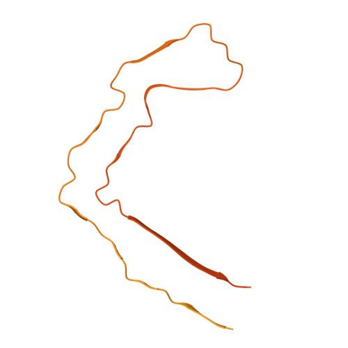
37VX | pdb_000037vx

CTE Type I structure

  • Classification: STRUCTURAL PROTEIN
  • Organism(s): Homo sapiens
  • Mutation(s): No 

  • Deposited: 2026-08-09 Released: 2026-10-14 
  • Deposition Author(s): Watanabe, R., Lee, E.B.
  • Funding Organization(s): National Institutes of Health/National Institute on Aging (NIH/NIA)

Experimental Data Snapshot

  • Method: ELECTRON MICROSCOPY
  • Resolution: 2.78 Å
  • Aggregation State: FILAMENT 
  • Reconstruction Method: HELICAL 

wwPDB Validation 3D Report Full Report

Validation slider image for 37VX

This is version 1.0 of the entry. See complete history. 

Re-refinement Note

This entry reflects an alternative modeling of the original data in: 6NWP

Literature

Structural heterogeneity of CTE Type I tau filaments with an interface-shifted polymorph.

Watanabe, R., Lee, E.B.

(2026) Biochem Biophys Res Commun 838: 154679-154679

  • DOI: https://doi.org/10.1016/j.bbrc.2026.154679
  • Primary Citation Related Structures: 
    37VX, 37VY

  • PubMed Abstract: 

    Tau filament types can differ in inter-protofilament packing while sharing the same protofilament fold. Motivated by our recent findings in vacuolar tauopathy, we analyzed cryo-electron microscopy (cryo-EM) data from brain-derived chronic traumatic encephalopathy (CTE) tau filaments deposited in EMPIAR-10313. We resolved the canonical CTE Type I structure and a previously unreported CTE Type I-like structure with a shifted protofilament interface at 2.78 and 2.96 Å, respectively. Mapping particle-state assignments back onto the original micrographs showed that the CTE Type I and CTE Type I-like packing states can occupy locally contiguous regions within the same fibrils. These findings identify an interface-shifted CTE Type I-like structure and support its local coexistence with CTE Type I within individual fibrils.


  • Organizational Affiliation: 
    • Translational Neuropathology Research Laboratory, Department of Pathology and Laboratory Medicine, Perelman School of Medicine at the University of Pennsylvania, 3400 Spruce St, Philadelphia, PA, 19104, USA; Department of Psychiatry, University of Tsukuba Hospital, 2-1-1 Amakubo, Tsukuba, Ibaraki, 305-8576, Japan. Electronic address: watanabe.ryohei.gb@u.tsukuba.ac.jp.

Macromolecule Content 

  • Total Structure Weight: 642.88 kDa 
  • Atom Count: 7,952 
  • Modeled Residue Count: 1,036 
  • Deposited Residue Count: 6,174 
  • Unique protein chains: 1

Macromolecules

Find similar proteins by:|  3D Structure
Entity ID: 1
MoleculeChains  Sequence LengthOrganismDetailsImage
Isoform Tau-F of Microtubule-associated protein tau441Homo sapiensMutation(s): 0 
UniProt & NIH Common Fund Data Resources
Find proteins for P10636 (Homo sapiens)
Go to UniProtKB:  P10636
PHAROS:  P10636
GTEx:  ENSG00000186868 
Entity Groups
Sequence Clusters30% Identity50% Identity70% Identity90% Identity95% Identity100% Identity
UniProt GroupP10636-8
Sequence Annotations
Expand
Reference Sequence

Experimental Data & Validation

Experimental Data

  • Method: ELECTRON MICROSCOPY
  • Resolution: 2.78 Å
  • Aggregation State: FILAMENT 
  • Reconstruction Method: HELICAL 
EM Software:
TaskSoftware PackageVersion
RECONSTRUCTIONRELION5.0.0

Structure Validation

View Full Validation Report


Entry History 

& Funding Information

Deposition Data


Funding OrganizationLocationGrant Number
National Institutes of Health/National Institute on Aging (NIH/NIA)United States--

Revision History  (Full details and data files)

  • Version 1.0: 2026-10-14
    Type: Initial release