38DC | pdb_000038dc

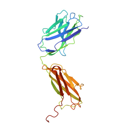
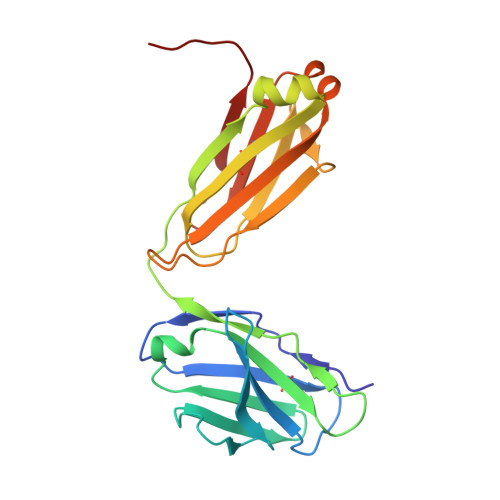
X-ray Structure of the FAB Fragment of a GluA2-specific Murine Monoclonal Antibody 15F1


Experimental Data Snapshot

  • Method: X-RAY DIFFRACTION
  • Resolution: 2.50 Å
  • R-Value Free: 
    0.257 (Depositor), 0.260 (DCC) 
  • R-Value Work: 
    0.209 (Depositor), 0.212 (DCC) 
  • R-Value Observed: 
    0.211 (Depositor) 

Starting Model: in silico
View more details

wwPDB Validation 3D Report Full Report

Validation slider image for 38DC

This is version 1.0 of the entry. See complete history. 

Literature

Programmable gold nanoparticle conjugates enable precise AMPA receptor localization at brain synapses by cryo-ET.

Spangler, C.J., Dhandapani, G., Matsui, A., Shiltseva, E.V., Nix, J., Pruneda, J.N., Elferich, J., Gouaux, E.

(2026) bioRxiv 

  • DOI: https://doi.org/10.64898/2026.09.18.752772
  • Primary Citation Related Structures: 
    38DC

  • PubMed Abstract: 

    Cryo-electron tomography (cryo-ET) is a powerful approach to image cells, organelles, viruses and molecular assemblies. However, resolving smaller complexes and molecules is challenging and presents a roadblock to the broad application of cryo-ET. Gold nanoparticles (AuNPs) have been used to localize otherwise unidentifiable molecules and complexes in tomograms but are typically tethered to the target of interest via a flexible linker. As a result, the position of the AuNP does not directly define the location of the target. Here we attach AuNPs to Fab antibody fragments via cysteine residues on defined elements of secondary structure and show that the AuNPs are rigidly bound to the Fab. We use these Fab-AuNP conjugates to locate AMPA receptors and probe their conformation upon desensitization with sub nanometer resolution in tomograms derived from brain slice lamella. By harnessing small, homogeneous AuNPs with precise conjugation to a Fab, we have developed reagents that enable direct investigation of conformational states of previously unidentifiable biomolecules and complexes in tomograms.


  • Organizational Affiliation: 
    • Vollum Institute, Oregon Health & Science University, Portland, OR 97239, USA.

Macromolecule Content 

  • Total Structure Weight: 97.77 kDa 
  • Atom Count: 6,912 
  • Modeled Residue Count: 886 
  • Deposited Residue Count: 898 
  • Unique protein chains: 2

Macromolecules

Find similar proteins by:|  3D Structure
Entity ID: 1
MoleculeChains  Sequence LengthOrganismDetailsImage
15F1 Fab heavy chain
A, C
233Spodoptera frugiperdaMutation(s): 0 
Entity Groups
Sequence Clusters30% Identity50% Identity70% Identity90% Identity95% Identity100% Identity
Sequence Annotations
Expand
Reference Sequence
Find similar proteins by:|  3D Structure
Entity ID: 2
MoleculeChains  Sequence LengthOrganismDetailsImage
15F1 Fab light chain
B, D
216Spodoptera frugiperdaMutation(s): 0 
Entity Groups
Sequence Clusters30% Identity50% Identity70% Identity90% Identity95% Identity100% Identity
Sequence Annotations
Expand
Reference Sequence

Experimental Data & Validation

Experimental Data

  • Method: X-RAY DIFFRACTION
  • Resolution: 2.50 Å
  • R-Value Free:  0.257 (Depositor), 0.260 (DCC) 
  • R-Value Work:  0.209 (Depositor), 0.212 (DCC) 
  • R-Value Observed: 0.211 (Depositor) 
Space Group: P 21 21 21
Unit Cell:
Length ( Å )Angle ( ˚ )
a = 62.461α = 90
b = 64.277β = 90
c = 204.89γ = 90
Software Package:
Software NamePurpose
PHENIXrefinement
Aimlessdata scaling
XDSdata reduction
PHENIXphasing
PDB_EXTRACTdata extraction

Structure Validation

View Full Validation Report


Entry History 

& Funding Information

Deposition Data


Funding OrganizationLocationGrant Number
Howard Hughes Medical Institute (HHMI)United States--
National Institutes of Health/National Institute of Neurological Disorders and Stroke (NIH/NINDS)United StatesR37NS038631

Revision History  (Full details and data files)

  • Version 1.0: 2026-11-04
    Type: Initial release