3A3J | pdb_00003a3j

Crystal structures of penicillin binding protein 5 from Haemophilus influenzae


Experimental Data Snapshot

  • Method: X-RAY DIFFRACTION
  • Resolution: 2.15 Å
  • R-Value Free: 
    0.269 (Depositor), 0.254 (DCC) 
  • R-Value Work: 
    0.208 (Depositor), 0.200 (DCC) 
  • R-Value Observed: 
    0.211 (Depositor) 

wwPDB Validation 3D Report Full Report

Validation slider image for 3A3J

This is version 1.3 of the entry. See complete history

Literature

Crystal structures of penicillin-binding proteins 4 and 5 from Haemophilus influenzae

Kawai, F.Clarke, T.B.Roper, D.I.Han, G.-J.Hwang, K.Y.Unzai, S.Obayashi, E.Park, S.-Y.Tame, J.R.H.

(2010) J Mol Biology 396: 634-645

  • DOI: https://doi.org/10.1016/j.jmb.2009.11.055
  • Primary Citation Related Structures: 
    3A3D, 3A3E, 3A3F, 3A3I, 3A3J

  • PubMed Abstract: 

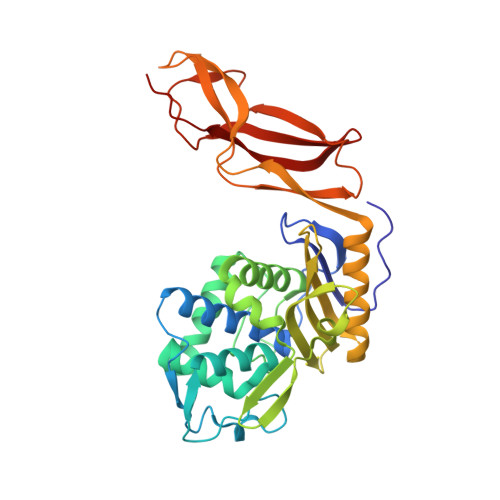
    We have determined high-resolution apo crystal structures of two low molecular weight penicillin-binding proteins (PBPs), PBP4 and PBP5, from Haemophilus influenzae, one of the most frequently found pathogens in the upper respiratory tract of children. Novel beta-lactams with notable antimicrobial activity have been designed, and crystal structures of PBP4 complexed with ampicillin and two of the novel molecules have also been determined. Comparing the apo form with those of the complexes, we find that the drugs disturb the PBP4 structure and weaken X-ray diffraction, to very different extents. PBP4 has recently been shown to act as a sensor of the presence of penicillins in Pseudomonas aeruginosa, and our models offer a clue to the structural basis for this effect. Covalently attached penicillins press against a phenylalanine residue near the active site and disturb the deacylation step. The ready inhibition of PBP4 by beta-lactams compared to PBP5 also appears to be related to the weaker interactions holding key residues in a catalytically competent position.


  • Organizational Affiliation
    • Yokohama City University, Suehiro 1-7-29, Tsurumi, Yokohama 230-0045, Japan.

Macromolecule Content 

  • Total Structure Weight: 38.09 kDa 
  • Atom Count: 2,793 
  • Modeled Residue Count: 344 
  • Deposited Residue Count: 344 
  • Unique protein chains: 1

Macromolecules

Find similar proteins by:|  3D Structure
Entity ID: 1
MoleculeChains  Sequence LengthOrganismDetailsImage
PBP5344Haemophilus influenzaeMutation(s): 0 
EC: 3.4.16.4
UniProt
Find proteins for P44466 (Haemophilus influenzae (strain ATCC 51907 / DSM 11121 / KW20 / Rd))
Explore P44466 
Go to UniProtKB:  P44466
Entity Groups
Sequence Clusters30% Identity50% Identity70% Identity90% Identity95% Identity100% Identity
UniProt GroupP44466
Sequence Annotations
Expand
Reference Sequence

Small Molecules

Ligands 1 Unique
IDChains Name / Formula / InChI Key2D Diagram3D Interactions
SO4

Query on SO4


Download:Ideal Coordinates CCD File
B [auth A]SULFATE ION
O4 S
QAOWNCQODCNURD-UHFFFAOYSA-L

Experimental Data & Validation

Experimental Data

  • Method: X-RAY DIFFRACTION
  • Resolution: 2.15 Å
  • R-Value Free:  0.269 (Depositor), 0.254 (DCC) 
  • R-Value Work:  0.208 (Depositor), 0.200 (DCC) 
  • R-Value Observed: 0.211 (Depositor) 
Space Group: P 21 21 21
Unit Cell:
Length ( Å )Angle ( ˚ )
a = 41.115α = 90
b = 52.694β = 90
c = 201.489γ = 90
Software Package:
Software NamePurpose
REFMACrefinement
HKL-2000data collection
HKL-2000data reduction
HKL-2000data scaling
MOLREPphasing

Structure Validation

View Full Validation Report


Entry History 

Deposition Data

Revision History  (Full details and data files)

  • Version 1.0: 2009-12-22
    Type: Initial release
  • Version 1.1: 2011-07-13
    Changes: Version format compliance
  • Version 1.2: 2013-11-20
    Changes: Database references
  • Version 1.3: 2024-03-13
    Changes: Data collection, Database references, Derived calculations