3AAU | pdb_00003aau

Bovine beta-trypsin bound to meta-diguanidino schiff base copper (II) chelate


Experimental Data Snapshot

  • Method: X-RAY DIFFRACTION
  • Resolution: 1.80 Å
  • R-Value Free: 
    0.202 (Depositor), 0.202 (DCC) 
  • R-Value Work: 
    0.173 (Depositor), 0.173 (DCC) 

Starting Model: experimental
View more details

wwPDB Validation 3D Report Full Report

Validation slider image for 3AAU

Ligand Structure Quality Assessment 


This is version 1.3 of the entry. See complete history

Literature

Structural basis for the design of novel Schiff base metal chelate inhibitors of trypsin

Iyaguchi, D.Kawano, S.Takada, K.Toyota, E.

(2010) Bioorg Med Chem 18: 2076-2080

  • DOI: https://doi.org/10.1016/j.bmc.2010.02.016
  • Primary Citation Related Structures: 
    3AAS, 3AAU, 3AAV

  • PubMed Abstract: 

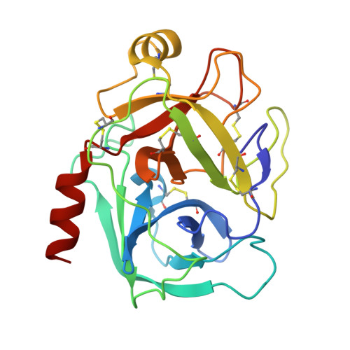
    The crystal structures of the complexes of bovine trypsin with m-guanidinosalicylidene-l-alaninato(aqua)copper(II) hydrochloride (inhibitor 1), [N,N'-bis(m-guanidinosalicylidene)ethylenediaminato]copper(II) (inhibitor 2), and [N,N'-bis(m-amidinosalicylidene)ethylenediaminato]copper(II) (inhibitor 4) have been determined. The guanidine-containing trypsin-inhibitors (1 and 2) bind to the trypsin active site in a manner similar to that previously reported for amidine-containing inhibitors, for example, m-amidinosalicylidene-l-alaninato(aqua)copper(II) hydrochloride (inhibitor 3). However, the binding mode of the guanidino groups of inhibitors 1 and 2 to Asp189 in the S1 pocket of trypsin was found to be markedly different from that of the amidino group of inhibitor 3. The present X-ray analyses revealed that the interactions of the metal ion of the inhibitors with the active site residues of trypsin play a crucial role in the binding affinity to the trypsin molecule. These structural information and inhibitory activity data for amidine- and guanidine-containing Schiff base metal chelate inhibitors provide new avenues for designing novel inhibitors against physiologically important trypsin-like serine proteases.


  • Organizational Affiliation
    • Faculty of Pharmaceutical Sciences, Health Sciences University of Hokkaido, Ishikari-Tobetsu, Hokkaido 061-0293, Japan.

Macromolecule Content 

  • Total Structure Weight: 23.81 kDa 
  • Atom Count: 1,918 
  • Modeled Residue Count: 223 
  • Deposited Residue Count: 223 
  • Unique protein chains: 1

Macromolecules

Find similar proteins by:|  3D Structure
Entity ID: 1
MoleculeChains  Sequence LengthOrganismDetailsImage
Cationic trypsin223Bos taurusMutation(s): 0 
EC: 3.4.21.4
UniProt
Find proteins for P00760 (Bos taurus)
Explore P00760 
Go to UniProtKB:  P00760
Entity Groups
Sequence Clusters30% Identity50% Identity70% Identity90% Identity95% Identity100% Identity
UniProt GroupP00760
Sequence Annotations
Expand
Reference Sequence

Small Molecules

Experimental Data & Validation

Experimental Data

  • Method: X-RAY DIFFRACTION
  • Resolution: 1.80 Å
  • R-Value Free:  0.202 (Depositor), 0.202 (DCC) 
  • R-Value Work:  0.173 (Depositor), 0.173 (DCC) 
Space Group: P 21 21 21
Unit Cell:
Length ( Å )Angle ( ˚ )
a = 54.183α = 90
b = 58.435β = 90
c = 66.498γ = 90
Software Package:
Software NamePurpose
CrystalCleardata collection
AMoREphasing
CNSrefinement
HKL-2000data reduction
HKL-2000data scaling

Structure Validation

View Full Validation Report


Ligand Structure Quality Assessment 


Entry History 

Deposition Data

Revision History  (Full details and data files)

  • Version 1.0: 2010-04-07
    Type: Initial release
  • Version 1.1: 2011-07-13
    Changes: Version format compliance
  • Version 1.2: 2023-11-01
    Changes: Data collection, Database references, Derived calculations, Refinement description
  • Version 1.3: 2024-10-30
    Changes: Structure summary