3C09 | pdb_00003c09

Crystal structure the Fab fragment of matuzumab (Fab72000) in complex with domain III of the extracellular region of EGFR


Experimental Data Snapshot

  • Method: X-RAY DIFFRACTION
  • Resolution: 3.20 Å
  • R-Value Free: 
    0.299 (Depositor), 0.299 (DCC) 
  • R-Value Work: 
    0.242 (Depositor), 0.240 (DCC) 
  • R-Value Observed: 
    0.245 (Depositor) 

Starting Models: experimental
View more details

wwPDB Validation 3D Report Full Report

Validation slider image for 3C09

Ligand Structure Quality Assessment 


This is version 1.6 of the entry. See complete history

Literature

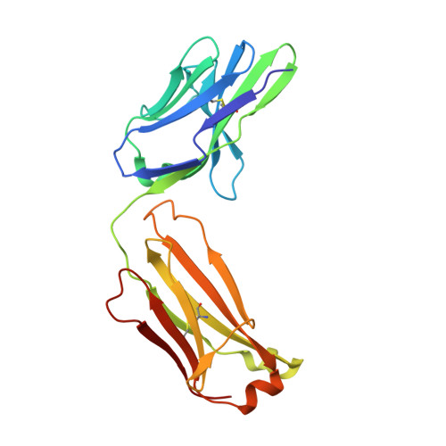
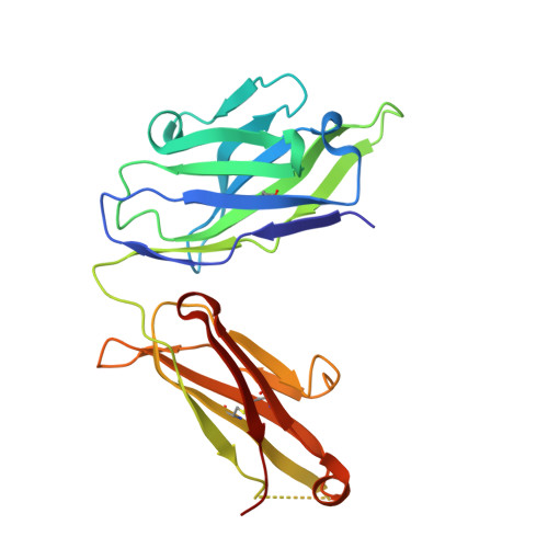
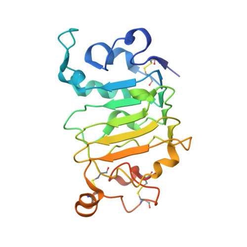
Matuzumab binding to EGFR prevents the conformational rearrangement required for dimerization.

Schmiedel, J.Blaukat, A.Li, S.Knochel, T.Ferguson, K.M.

(2008) Cancer Cell 13: 365-373

  • DOI: https://doi.org/10.1016/j.ccr.2008.02.019
  • Primary Citation Related Structures: 
    3C08, 3C09

  • PubMed Abstract: 

    An increasing number of therapeutic antibodies targeting tumors that express the epidermal growth factor receptor (EGFR) are in clinical use or late stages of clinical development. Here we investigate the molecular basis for inhibition of EGFR activation by the therapeutic antibody matuzumab (EMD72000). We describe the X-ray crystal structure of the Fab fragment of matuzumab (Fab72000) in complex with isolated domain III from the extracellular region of EGFR. Fab72000 interacts with an epitope on EGFR that is distinct from the ligand-binding region on domain III and from the cetuximab/Erbitux epitope. Matuzumab blocks ligand-induced receptor activation indirectly by sterically preventing the domain rearrangement and local conformational changes that must occur for high-affinity ligand binding and receptor dimerization.


  • Organizational Affiliation
    • Department of Physiology, University of Pennsylvania School of Medicine, Philadelphia, PA 19104, USA.

Macromolecule Content 

  • Total Structure Weight: 144.88 kDa 
  • Atom Count: 8,532 
  • Modeled Residue Count: 1,175 
  • Deposited Residue Count: 1,298 
  • Unique protein chains: 3

Macromolecules

Find similar proteins by:|  3D Structure
Entity ID: 1
MoleculeChains  Sequence LengthOrganismDetailsImage
Matuzumab Fab Light chainA [auth L],
D [auth B]
212Mus musculusMutation(s): 0 
UniProt & NIH Common Fund Data Resources
Find proteins for P01834 (Homo sapiens)
Explore P01834 
Go to UniProtKB:  P01834
PHAROS:  P01834
Entity Groups
Sequence Clusters30% Identity50% Identity70% Identity90% Identity95% Identity100% Identity
UniProt GroupP01834
Sequence Annotations
Expand
Reference Sequence
Find similar proteins by:|  3D Structure
Entity ID: 2
MoleculeChains  Sequence LengthOrganismDetailsImage
Matuzumab Fab Heavy chainB [auth H],
E [auth C]
223Mus musculusMutation(s): 0 
UniProt & NIH Common Fund Data Resources
Find proteins for P01857 (Homo sapiens)
Explore P01857 
Go to UniProtKB:  P01857
PHAROS:  P01857
GTEx:  ENSG00000211896 
Entity Groups
Sequence Clusters30% Identity50% Identity70% Identity90% Identity95% Identity100% Identity
UniProt GroupP01857
Sequence Annotations
Expand
Reference Sequence
Find similar proteins by:|  3D Structure
Entity ID: 3
MoleculeChains  Sequence LengthOrganismDetailsImage
Epidermal growth factor receptorC [auth A],
F [auth D]
214Homo sapiensMutation(s): 0 
Gene Names: EGFR
EC: 2.7.10.1
UniProt & NIH Common Fund Data Resources
Find proteins for P00533 (Homo sapiens)
Explore P00533 
Go to UniProtKB:  P00533
PHAROS:  P00533
GTEx:  ENSG00000146648 
Entity Groups
Sequence Clusters30% Identity50% Identity70% Identity90% Identity95% Identity100% Identity
UniProt GroupP00533
Sequence Annotations
Expand
Reference Sequence

Small Molecules

Ligands 3 Unique
IDChains Name / Formula / InChI Key2D Diagram3D Interactions
NAG

Query on NAG


Download:Ideal Coordinates CCD File
G [auth A]
H [auth A]
K [auth A]
L [auth A]
M [auth A]
G [auth A],
H [auth A],
K [auth A],
L [auth A],
M [auth A],
N [auth A],
O [auth D],
P [auth D],
R [auth D],
S [auth D]
2-acetamido-2-deoxy-beta-D-glucopyranose
C8 H15 N O6
OVRNDRQMDRJTHS-FMDGEEDCSA-N
MAN

Query on MAN


Download:Ideal Coordinates CCD File
J [auth A]alpha-D-mannopyranose
C6 H12 O6
WQZGKKKJIJFFOK-PQMKYFCFSA-N
BMA

Query on BMA


Download:Ideal Coordinates CCD File
I [auth A],
Q [auth D]
beta-D-mannopyranose
C6 H12 O6
WQZGKKKJIJFFOK-RWOPYEJCSA-N

Experimental Data & Validation

Experimental Data

  • Method: X-RAY DIFFRACTION
  • Resolution: 3.20 Å
  • R-Value Free:  0.299 (Depositor), 0.299 (DCC) 
  • R-Value Work:  0.242 (Depositor), 0.240 (DCC) 
  • R-Value Observed: 0.245 (Depositor) 
Space Group: C 1 2 1
Unit Cell:
Length ( Å )Angle ( ˚ )
a = 141.073α = 90
b = 205.035β = 117.49
c = 81.577γ = 90
Software Package:
Software NamePurpose
DENZOdata reduction
SCALEPACKdata scaling
PHASERphasing
REFMACrefinement
PDB_EXTRACTdata extraction

Structure Validation

View Full Validation Report


Ligand Structure Quality Assessment 


Entry History 

Deposition Data

Revision History  (Full details and data files)

  • Version 1.0: 2008-04-15
    Type: Initial release
  • Version 1.1: 2011-07-13
    Changes: Non-polymer description, Version format compliance
  • Version 1.2: 2017-10-25
    Changes: Refinement description
  • Version 1.3: 2019-12-25
    Changes: Database references, Refinement description, Source and taxonomy
  • Version 1.4: 2020-07-29
    Type: Remediation
    Reason: Carbohydrate remediation
    Changes: Data collection, Derived calculations, Structure summary
  • Version 1.5: 2023-11-01
    Changes: Data collection, Database references, Refinement description, Structure summary
  • Version 1.6: 2024-11-13
    Changes: Source and taxonomy, Structure summary