3CQD | pdb_00003cqd

Structure of the tetrameric inhibited form of phosphofructokinase-2 from Escherichia coli


Experimental Data Snapshot

  • Method: X-RAY DIFFRACTION
  • Resolution: 1.98 Å
  • R-Value Free: 
    0.234 (Depositor), 0.233 (DCC) 
  • R-Value Work: 
    0.186 (Depositor), 0.185 (DCC) 
  • R-Value Observed: 
    0.189 (Depositor) 

Starting Model: experimental
View more details

wwPDB Validation 3D Report Full Report

Validation slider image for 3CQD

Ligand Structure Quality Assessment 


This is version 1.3 of the entry. See complete history

Literature

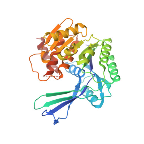
Crystallographic structure of phosphofructokinase-2 from Escherichia coli in complex with two ATP molecules. Implications for substrate inhibition.

Cabrera, R.Ambrosio, A.L.Garratt, R.C.Guixe, V.Babul, J.

(2008) J Mol Biology 383: 588-602

  • DOI: https://doi.org/10.1016/j.jmb.2008.08.029
  • Primary Citation Related Structures: 
    3CQD

  • PubMed Abstract: 

    Phosphofructokinase-1 and -2 (Pfk-1 and Pfk-2, respectively) from Escherichia coli belong to different homologous superfamilies. However, in spite of the lack of a common ancestor, they share the ability to catalyze the same reaction and are inhibited by the substrate MgATP. Pfk-2, an ATP-dependent 6-phosphofructokinase member of the ribokinase-like superfamily, is a homodimer of 66 kDa subunits whose oligomerization state is necessary for catalysis and stability. The presence of MgATP favors the tetrameric form of the enzyme. In this work, we describe the structure of Pfk-2 in its inhibited tetrameric form, with each subunit bound to two ATP molecules and two Mg ions. The present structure indicates that substrate inhibition occurs due to the sequential binding of two MgATP molecules per subunit, the first at the usual site occupied by the nucleotide in homologous enzymes and the second at the allosteric site, making a number of direct and Mg-mediated interactions with the first. Two configurations are observed for the second MgATP, one of which involves interactions with Tyr23 from the adjacent subunit in the dimer and the other making an unusual non-Watson-Crick base pairing with the adenine in the substrate ATP. The oligomeric state observed in the crystal is tetrameric, and some of the structural elements involved in the binding of the substrate and allosteric ATPs are also participating in the dimer-dimer interface. This structure also provides the grounds to compare analogous features of the nonhomologous phosphofructokinases from E. coli.


  • Organizational Affiliation
    • Departamento de Biología, Facultad de Ciencias, Universidad de Chile, Casilla 653, Santiago, Chile.

Macromolecule Content 

  • Total Structure Weight: 67.1 kDa 
  • Atom Count: 5,126 
  • Modeled Residue Count: 612 
  • Deposited Residue Count: 618 
  • Unique protein chains: 1

Macromolecules

Find similar proteins by:|  3D Structure
Entity ID: 1
MoleculeChains  Sequence LengthOrganismDetailsImage
6-phosphofructokinase isozyme 2
A, B
309Escherichia coliMutation(s): 0 
EC: 2.7.1.11
UniProt
Find proteins for P06999 (Escherichia coli (strain K12))
Explore P06999 
Go to UniProtKB:  P06999
Entity Groups
Sequence Clusters30% Identity50% Identity70% Identity90% Identity95% Identity100% Identity
UniProt GroupP06999
Sequence Annotations
Expand
Reference Sequence

Small Molecules

Ligands 2 Unique
IDChains Name / Formula / InChI Key2D Diagram3D Interactions
ATP

Query on ATP


Download:Ideal Coordinates CCD File
F [auth A],
G [auth A],
I [auth B],
J [auth B]
ADENOSINE-5'-TRIPHOSPHATE
C10 H16 N5 O13 P3
ZKHQWZAMYRWXGA-KQYNXXCUSA-N
MG

Query on MG


Download:Ideal Coordinates CCD File
C [auth A],
D [auth A],
E [auth A],
H [auth B]
MAGNESIUM ION
Mg
JLVVSXFLKOJNIY-UHFFFAOYSA-N

Experimental Data & Validation

Experimental Data

  • Method: X-RAY DIFFRACTION
  • Resolution: 1.98 Å
  • R-Value Free:  0.234 (Depositor), 0.233 (DCC) 
  • R-Value Work:  0.186 (Depositor), 0.185 (DCC) 
  • R-Value Observed: 0.189 (Depositor) 
Space Group: P 2 2 21
Unit Cell:
Length ( Å )Angle ( ˚ )
a = 42.822α = 90
b = 86.834β = 90
c = 171.308γ = 90
Software Package:
Software NamePurpose
REFMACrefinement
MAR345data collection
MOSFLMdata reduction
SCALAdata scaling
PHASERphasing

Structure Validation

View Full Validation Report


Ligand Structure Quality Assessment 


Entry History 

Deposition Data

Revision History  (Full details and data files)

  • Version 1.0: 2008-09-16
    Type: Initial release
  • Version 1.1: 2011-07-13
    Changes: Version format compliance
  • Version 1.2: 2017-10-25
    Changes: Refinement description
  • Version 1.3: 2023-08-30
    Changes: Data collection, Database references, Derived calculations, Refinement description