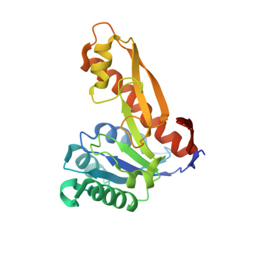
3DCJ

Crystal structure of glycinamide formyltransferase (PurN) from Mycobacterium tuberculosis in complex with 5-methyl-5,6,7,8-tetrahydrofolic acid derivative


Experimental Data Snapshot

  • Method: X-RAY DIFFRACTION
  • Resolution: 2.20 Å
  • R-Value Free: 0.243 
  • R-Value Work: 0.184 
  • R-Value Observed: 0.187 

Starting Model: experimental
View more details

wwPDB Validation   3D Report Full Report


Ligand Structure Quality Assessment 


This is version 1.3 of the entry. See complete history


Literature

Structures of glycinamide ribonucleotide transformylase (PurN) from Mycobacterium tuberculosis reveal a novel dimer with relevance to drug discovery.

Zhang, Z.Caradoc-Davies, T.T.Dickson, J.M.Baker, E.N.Squire, C.J.

(2009) J Mol Biol 389: 722-733

  • DOI: https://doi.org/10.1016/j.jmb.2009.04.044
  • Primary Citation of Related Structures:  
    3DA8, 3DCJ

  • PubMed Abstract: 

    Enzymes from the de novo purine biosynthetic pathway have been exploited for the development of anti-cancer drugs, and represent novel targets for anti-bacterial drug development. In Mycobacterium tuberculosis, the cause of tuberculosis, this pathway has been identified as essential for growth and survival. The structure of M. tuberculosis PurN (MtPurN) has been determined in complex with magnesium and iodide at 1.30 A resolution, and with cofactor analogue, 5-methyltetrahydrofolate (5MTHF) at 2.2 A resolution. The structure shows a Rossmann-type fold that is very similar to the known structures of the human and E. coli PurN proteins. In contrast, MtPurN forms a dimer that is quite different from that formed by the Escherichia coli PurN, and which suggests a mechanism whereby communication could take place between the two active sites. Differences are seen in two active site loops and in the binding mode of the 5MTHF cofactor analogue between the two MtPurN molecules of the dimer. A binding site for halide ions is found in the dimer interface, and bound magnesium and iodide ions in the active site suggest sites that might be exploited in potential drug discovery strategies.


  • Organizational Affiliation

    School of Biological Sciences & Maurice Wilkins Centre for Molecular Biodiscovery, University of Auckland, Private Bag 92010, Auckland 1142, New Zealand.


Macromolecules
Find similar proteins by:  (by identity cutoff)  |  3D Structure
Entity ID: 1
MoleculeChains Sequence LengthOrganismDetailsImage
Probable 5'-phosphoribosylglycinamide formyltransferase purN
A, B
215Mycobacterium tuberculosisMutation(s): 0 
Gene Names: purNMT0983Rv0956
EC: 2.1.2.2
UniProt
Find proteins for P9WHM5 (Mycobacterium tuberculosis (strain ATCC 25618 / H37Rv))
Explore P9WHM5 
Go to UniProtKB:  P9WHM5
Entity Groups  
Sequence Clusters30% Identity50% Identity70% Identity90% Identity95% Identity100% Identity
UniProt GroupP9WHM5
Sequence Annotations
Expand
  • Reference Sequence
Small Molecules
Ligands 4 Unique
IDChains Name / Formula / InChI Key2D Diagram3D Interactions
THH
Query on THH

Download Ideal Coordinates CCD File 
C [auth A],
F [auth B]
N-[4-({[(6S)-2-AMINO-4-HYDROXY-5-METHYL-5,6,7,8-TETRAHYDROPTERIDIN-6-YL]METHYL}AMINO)BENZOYL]-L-GLUTAMIC ACID
C20 H25 N7 O6
ZNOVTXRBGFNYRX-STQMWFEESA-N
NO3
Query on NO3

Download Ideal Coordinates CCD File 
G [auth B]NITRATE ION
N O3
NHNBFGGVMKEFGY-UHFFFAOYSA-N
CL
Query on CL

Download Ideal Coordinates CCD File 
E [auth A],
H [auth B]
CHLORIDE ION
Cl
VEXZGXHMUGYJMC-UHFFFAOYSA-M
MG
Query on MG

Download Ideal Coordinates CCD File 
D [auth A]MAGNESIUM ION
Mg
JLVVSXFLKOJNIY-UHFFFAOYSA-N
Experimental Data & Validation

Experimental Data

  • Method: X-RAY DIFFRACTION
  • Resolution: 2.20 Å
  • R-Value Free: 0.243 
  • R-Value Work: 0.184 
  • R-Value Observed: 0.187 
  • Space Group: P 1 21 1
Unit Cell:
Length ( Å )Angle ( ˚ )
a = 62.153α = 90
b = 61.66β = 103.31
c = 61.525γ = 90
Software Package:
Software NamePurpose
REFMACrefinement
MAR345dtbdata collection
MOSFLMdata reduction
SCALAdata scaling
PHASERphasing
PHENIXrefinement

Structure Validation

View Full Validation Report


Ligand Structure Quality Assessment 


Entry History 

Deposition Data

Revision History  (Full details and data files)

  • Version 1.0: 2009-05-12
    Type: Initial release
  • Version 1.1: 2011-07-13
    Changes: Version format compliance
  • Version 1.2: 2019-07-24
    Changes: Data collection, Refinement description
  • Version 1.3: 2023-08-30
    Changes: Data collection, Database references, Derived calculations, Refinement description