3DLA

X-ray crystal structure of glutamine-dependent NAD+ synthetase from Mycobacterium tuberculosis bound to NaAD+ and DON


Experimental Data Snapshot

  • Method: X-RAY DIFFRACTION
  • Resolution: 2.35 Å
  • R-Value Free: 
    0.246 (Depositor) 
  • R-Value Work: 
    0.184 (Depositor) 
  • R-Value Observed: 
    0.187 (Depositor) 

wwPDB Validation   3D Report Full Report


Ligand Structure Quality Assessment 

Created with Raphaël 2.3.0Worse 01 BetterLigand structure goodness of fit to experimental dataBest fitted DNDClick on this verticalbar to view details

This is version 2.0 of the entry. See complete history


Literature

Regulation of active site coupling in glutamine-dependent NAD(+) synthetase.

LaRonde-LeBlanc, N.Resto, M.Gerratana, B.

(2009) Nat Struct Mol Biol 16: 421-429

  • DOI: https://doi.org/10.1038/nsmb.1567
  • Primary Citation of Related Structures:  
    3DLA

  • PubMed Abstract: 

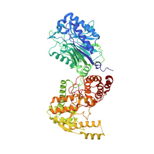
    NAD(+) is an essential metabolite both as a cofactor in energy metabolism and redox homeostasis and as a regulator of cellular processes. In contrast to humans, Mycobacterium tuberculosis NAD(+) biosynthesis is absolutely dependent on the activity of a multifunctional glutamine-dependent NAD(+) synthetase, which catalyzes the ATP-dependent formation of NAD(+) at the synthetase domain using ammonia derived from L-glutamine in the glutaminase domain. Here we report the kinetics and structural characterization of M. tuberculosis NAD(+) synthetase. The kinetics data strongly suggest tightly coupled regulation of the catalytic activities. The structure, the first of a glutamine-dependent NAD(+) synthetase, reveals a homooctameric subunit organization suggesting a tight dependence of catalysis on the quaternary structure, a 40-A intersubunit ammonia tunnel and structural elements that may be involved in the transfer of information between catalytic sites.


  • Organizational Affiliation

    Department of Chemistry and Biochemistry, University of Maryland, College Park, Maryland 20742-2021, USA. nlaronde@umd.edu


Macromolecules
Find similar proteins by:  (by identity cutoff)  |  3D Structure
Entity ID: 1
MoleculeChains Sequence LengthOrganismDetailsImage
Glutamine-dependent NAD(+) synthetase
A, B, C, D
680Mycobacterium tuberculosisMutation(s): 0 
Gene Names: nadERv2438cMT2513MTCY428.08
EC: 6.3.5.1
UniProt
Find proteins for P9WJJ3 (Mycobacterium tuberculosis (strain ATCC 25618 / H37Rv))
Explore P9WJJ3 
Go to UniProtKB:  P9WJJ3
Entity Groups  
Sequence Clusters30% Identity50% Identity70% Identity90% Identity95% Identity100% Identity
UniProt GroupP9WJJ3
Sequence Annotations
Expand
  • Reference Sequence
Small Molecules
Ligands 3 Unique
IDChains Name / Formula / InChI Key2D Diagram3D Interactions
DND
Query on DND

Download Ideal Coordinates CCD File 
E [auth A],
L [auth B],
Q [auth C],
V [auth D]
NICOTINIC ACID ADENINE DINUCLEOTIDE
C21 H27 N6 O15 P2
SENPVEZBRZQVST-HISDBWNOSA-O
ONL
Query on ONL

Download Ideal Coordinates CCD File 
F [auth A],
G [auth A],
H [auth A],
I [auth A]
5-OXO-L-NORLEUCINE
C6 H11 N O3
KSIJECNNZVKMJG-YFKPBYRVSA-N
GOL
Query on GOL

Download Ideal Coordinates CCD File 
AA [auth D]
J [auth A]
K [auth A]
M [auth B]
N [auth B]
AA [auth D],
J [auth A],
K [auth A],
M [auth B],
N [auth B],
O [auth B],
P [auth B],
R [auth C],
S [auth C],
T [auth C],
U [auth C],
W [auth D],
X [auth D],
Y [auth D],
Z [auth D]
GLYCEROL
C3 H8 O3
PEDCQBHIVMGVHV-UHFFFAOYSA-N
Experimental Data & Validation

Experimental Data

  • Method: X-RAY DIFFRACTION
  • Resolution: 2.35 Å
  • R-Value Free:  0.246 (Depositor) 
  • R-Value Work:  0.184 (Depositor) 
  • R-Value Observed: 0.187 (Depositor) 
Space Group: P 41 21 2
Unit Cell:
Length ( Å )Angle ( ˚ )
a = 178.13α = 90
b = 178.13β = 90
c = 215.177γ = 90
Software Package:
Software NamePurpose
REFMACrefinement
HKL-2000data reduction
HKL-2000data scaling
SOLVEphasing

Structure Validation

View Full Validation Report


Ligand Structure Quality Assessment 

Created with Raphaël 2.3.0Worse 01 BetterLigand structure goodness of fit to experimental dataBest fitted DNDClick on this verticalbar to view details

Entry History 

Deposition Data

Revision History  (Full details and data files)

  • Version 1.0: 2009-03-10
    Type: Initial release
  • Version 1.1: 2011-07-13
    Changes: Advisory, Refinement description, Version format compliance
  • Version 2.0: 2019-10-23
    Changes: Advisory, Atomic model, Data collection, Database references, Derived calculations, Non-polymer description, Structure summary