3DLS | pdb_00003dls

Crystal structure of human PAS kinase bound to ADP


Experimental Data Snapshot

  • Method: X-RAY DIFFRACTION
  • Resolution: 2.30 Å
  • R-Value Free: 
    0.297 (Depositor), 0.295 (DCC) 
  • R-Value Work: 
    0.241 (Depositor), 0.240 (DCC) 
  • R-Value Observed: 
    0.244 (Depositor) 

wwPDB Validation 3D Report Full Report

Validation slider image for 3DLS

Ligand Structure Quality Assessment 


This is version 1.5 of the entry. See complete history

Literature

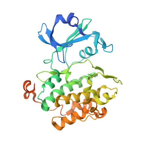
Structural bases of PAS domain-regulated kinase (PASK) activation in the absence of activation loop phosphorylation.

Kikani, C.K.Antonysamy, S.A.Bonanno, J.B.Romero, R.Zhang, F.F.Russell, M.Gheyi, T.Iizuka, M.Emtage, S.Sauder, J.M.Turk, B.E.Burley, S.K.Rutter, J.

(2010) J Biological Chem 285: 41034-41043

  • DOI: https://doi.org/10.1074/jbc.M110.157594
  • Primary Citation Related Structures: 
    3DLS

  • PubMed Abstract: 

    Per-Arnt-Sim (PAS) domain-containing protein kinase (PASK) is an evolutionary conserved protein kinase that coordinates cellular metabolism with metabolic demand in yeast and mammals. The molecular mechanisms underlying PASK regulation, however, remain unknown. Herein, we describe a crystal structure of the kinase domain of human PASK, which provides insights into the regulatory mechanisms governing catalysis. We show that the kinase domain adopts an active conformation and has catalytic activity in vivo and in vitro in the absence of activation loop phosphorylation. Using site-directed mutagenesis and structural comparison with active and inactive kinases, we identified several key structural features in PASK that enable activation loop phosphorylation-independent activity. Finally, we used combinatorial peptide library screening to determine that PASK prefers basic residues at the P-3 and P-5 positions in substrate peptides. Our results describe the key features of the PASK structure and how those features are important for PASK activity and substrate selection.


  • Organizational Affiliation
    • Department of Biochemistry, University of Utah School of Medicine, Salt Lake City, Utah 84112-5650, USA.

Macromolecule Content 

  • Total Structure Weight: 230.28 kDa 
  • Atom Count: 13,724 
  • Modeled Residue Count: 1,693 
  • Deposited Residue Count: 2,010 
  • Unique protein chains: 1

Macromolecules

Find similar proteins by:|  3D Structure
Entity ID: 1
MoleculeChains  Sequence LengthOrganismDetailsImage
PAS domain-containing serine/threonine-protein kinase
A, B, C, D, E
A, B, C, D, E, F
335Homo sapiensMutation(s): 0 
Gene Names: PASKKIAA0135
EC: 2.7.11.1
UniProt & NIH Common Fund Data Resources
Find proteins for Q96RG2 (Homo sapiens)
Explore Q96RG2 
Go to UniProtKB:  Q96RG2
PHAROS:  Q96RG2
GTEx:  ENSG00000115687 
Entity Groups
Sequence Clusters30% Identity50% Identity70% Identity90% Identity95% Identity100% Identity
UniProt GroupQ96RG2
Sequence Annotations
Expand
Reference Sequence

Small Molecules

Ligands 2 Unique
IDChains Name / Formula / InChI Key2D Diagram3D Interactions
ADP

Query on ADP


Download:Ideal Coordinates CCD File
BA [auth E]
EA [auth F]
K [auth A]
P [auth B]
U [auth C]
BA [auth E],
EA [auth F],
K [auth A],
P [auth B],
U [auth C],
Y [auth D]
ADENOSINE-5'-DIPHOSPHATE
C10 H15 N5 O10 P2
XTWYTFMLZFPYCI-KQYNXXCUSA-N
MG

Query on MG


Download:Ideal Coordinates CCD File
AA [auth E]
CA [auth F]
DA [auth F]
G [auth A]
H [auth A]
AA [auth E],
CA [auth F],
DA [auth F],
G [auth A],
H [auth A],
I [auth A],
J [auth A],
L [auth B],
M [auth B],
N [auth B],
O [auth B],
Q [auth C],
R [auth C],
S [auth C],
T [auth C],
V [auth D],
W [auth D],
X [auth D],
Z [auth E]
MAGNESIUM ION
Mg
JLVVSXFLKOJNIY-UHFFFAOYSA-N

Experimental Data & Validation

Experimental Data

  • Method: X-RAY DIFFRACTION
  • Resolution: 2.30 Å
  • R-Value Free:  0.297 (Depositor), 0.295 (DCC) 
  • R-Value Work:  0.241 (Depositor), 0.240 (DCC) 
  • R-Value Observed: 0.244 (Depositor) 
Space Group: P 1
Unit Cell:
Length ( Å )Angle ( ˚ )
a = 85.778α = 77.28
b = 85.84β = 77.5
c = 94.154γ = 60.09
Software Package:
Software NamePurpose
REFMACrefinement
PDB_EXTRACTdata extraction

Structure Validation

View Full Validation Report


Ligand Structure Quality Assessment 


Entry History 

Revision History  (Full details and data files)

  • Version 1.0: 2008-08-26
    Type: Initial release
  • Version 1.1: 2011-07-13
    Changes: Derived calculations, Version format compliance
  • Version 1.2: 2015-08-19
    Changes: Database references
  • Version 1.3: 2018-11-14
    Changes: Data collection, Structure summary
  • Version 1.4: 2021-02-10
    Changes: Database references, Derived calculations, Structure summary
  • Version 1.5: 2024-02-21
    Changes: Data collection, Database references