


Crystal Structure of Rrna-Methylase from Clostridium Thermocellum
Patskovsky, Y., Ramagopal, U.A., Toro, R., Rutter, M., Hu, S., Bain, K., Sauder, J.M., Burley, S.K., Almo, S.C.To be published.
Experimental Data Snapshot
Entity ID: 1 | |||||
|---|---|---|---|---|---|
| Molecule | Chains | Sequence Length | Organism | Details | Image |
| Putative rRNA methylase | 197 | Acetivibrio thermocellus ATCC 27405 | Mutation(s): 0 Gene Names: Cthe_0696 | 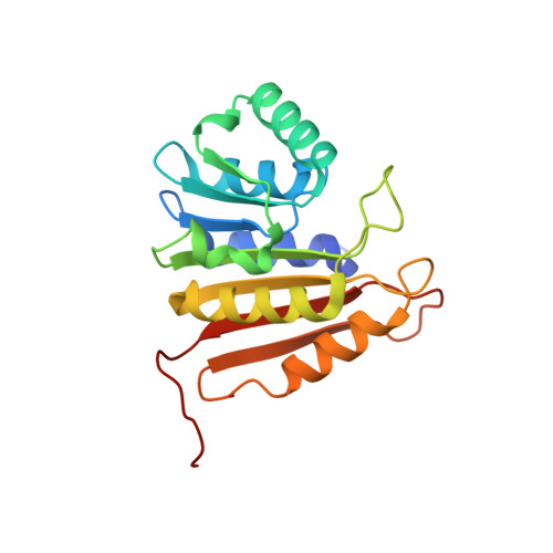 | |
UniProt | |||||
Find proteins for A3DDA2 (Acetivibrio thermocellus (strain ATCC 27405 / DSM 1237 / JCM 9322 / NBRC 103400 / NCIMB 10682 / NRRL B-4536 / VPI 7372)) Explore A3DDA2 Go to UniProtKB: A3DDA2 | |||||
Entity Groups | |||||
| Sequence Clusters | 30% Identity50% Identity70% Identity90% Identity95% Identity100% Identity | ||||
| UniProt Group | A3DDA2 | ||||
Sequence AnnotationsExpand | |||||
| |||||
| Ligands 3 Unique | |||||
|---|---|---|---|---|---|
| ID | Chains | Name / Formula / InChI Key | 2D Diagram | 3D Interactions | |
| SAM Query on SAM | AA [auth I] BA [auth J] K [auth A] P [auth B] S [auth C] | S-ADENOSYLMETHIONINE C15 H22 N6 O5 S MEFKEPWMEQBLKI-FCKMPRQPSA-N |  | ||
| SO4 Query on SO4 | L [auth A], M [auth A], N [auth A], Q [auth B], T [auth C] | SULFATE ION O4 S QAOWNCQODCNURD-UHFFFAOYSA-L |  | ||
| GOL Query on GOL | O [auth A], R [auth B], X [auth F], Z [auth H] | GLYCEROL C3 H8 O3 PEDCQBHIVMGVHV-UHFFFAOYSA-N |  | ||
| Length ( Å ) | Angle ( ˚ ) |
|---|---|
| a = 70.156 | α = 90 |
| b = 126.913 | β = 99.54 |
| c = 125.611 | γ = 90 |
| Software Name | Purpose |
|---|---|
| SHELX | model building |
| REFMAC | refinement |
| HKL-2000 | data reduction |
| HKL-2000 | data scaling |
| SHELX | phasing |


