


Structure and mechanism of a new family of prokaryotic nucleoside diphosphatases.
Proudfoot, M., Brown, M., Singer, A.U., Jobin, M.-C., Xu, X., Zheng, H., Chang, C., Edwards, A.M., Joachimiak, A., Savchenko, A., Yakunin, A.F.To be published.
Experimental Data Snapshot
Starting Model: experimental
View more details
Entity ID: 1 | |||||
|---|---|---|---|---|---|
| Molecule | Chains | Sequence Length | Organism | Details | Image |
| Phosphatase SC4828 | 237 | Streptomyces coelicolor A3(2) | Mutation(s): 0 Gene Names: SCO5041, SCK7.14, SCK7.14c EC: 3.6.1.6 | 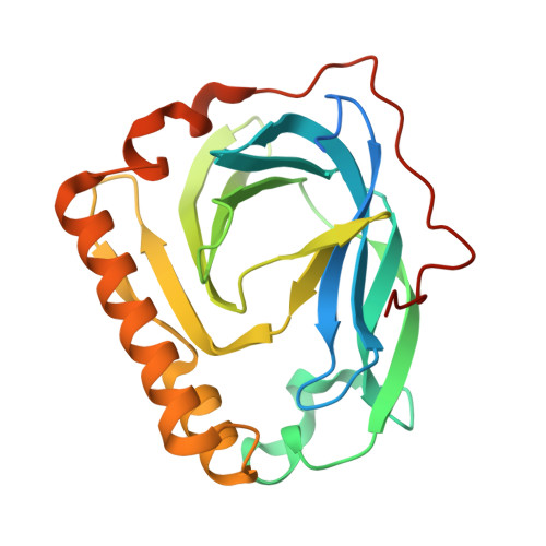 | |
UniProt | |||||
Entity Groups | |||||
| Sequence Clusters | 30% Identity50% Identity70% Identity90% Identity95% Identity100% Identity | ||||
| UniProt Group | Q9FBN7 | ||||
Sequence AnnotationsExpand | |||||
Reference Sequence | |||||
| Ligands 4 Unique | |||||
|---|---|---|---|---|---|
| ID | Chains | Name / Formula / InChI Key | 2D Diagram | 3D Interactions | |
| GP2 Download:Ideal Coordinates CCD File | G [auth A] | PHOSPHOMETHYLPHOSPHONIC ACID GUANOSYL ESTER C11 H17 N5 O10 P2 OCJWYBKRHNXUME-KQYNXXCUSA-N |  | ||
| GOL Download:Ideal Coordinates CCD File | H [auth A] | GLYCEROL C3 H8 O3 PEDCQBHIVMGVHV-UHFFFAOYSA-N |  | ||
| CA Download:Ideal Coordinates CCD File | B [auth A], C [auth A], D [auth A] | CALCIUM ION Ca BHPQYMZQTOCNFJ-UHFFFAOYSA-N |  | ||
| NA Download:Ideal Coordinates CCD File | E [auth A], F [auth A] | SODIUM ION Na FKNQFGJONOIPTF-UHFFFAOYSA-N |  | ||
| Length ( Å ) | Angle ( ˚ ) |
|---|---|
| a = 36.941 | α = 90 |
| b = 79.706 | β = 90 |
| c = 82.507 | γ = 90 |
| Software Name | Purpose |
|---|---|
| CrystalClear | data collection |
| PHASER | phasing |
| REFMAC | refinement |
| HKL-2000 | data reduction |
| HKL-2000 | data scaling |


