3EYH

Crystal structures of JAK1 and JAK2 inhibitor complexes


Experimental Data Snapshot

  • Method: X-RAY DIFFRACTION
  • Resolution: 2.00 Å
  • R-Value Free: 
    0.237 (Depositor), 0.240 (DCC) 
  • R-Value Work: 
    0.174 (Depositor), 0.180 (DCC) 
  • R-Value Observed: 
    0.178 (Depositor) 

wwPDB Validation   3D Report Full Report


Ligand Structure Quality Assessment 

Created with Raphaël 2.3.0Worse 01 BetterLigand structure goodness of fit to experimental dataBest fitted IZAClick on this verticalbar to view details

This is version 1.3 of the entry. See complete history


Literature

Dissecting specificity in the Janus kinases: the structures of JAK-specific inhibitors complexed to the JAK1 and JAK2 protein tyrosine kinase domains.

Williams, N.K.Bamert, R.S.Patel, O.Wang, C.Walden, P.M.Wilks, A.F.Fantino, E.Rossjohn, J.Lucet, I.S.

(2009) J Mol Biology 387: 219-232

  • DOI: https://doi.org/10.1016/j.jmb.2009.01.041
  • Primary Citation of Related Structures:  
    3EYG, 3EYH, 3FUP

  • PubMed Abstract: 

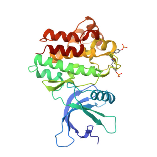
    The Janus kinases (JAKs) are a pivotal family of protein tyrosine kinases (PTKs) that play prominent roles in numerous cytokine signaling pathways, with aberrant JAK activity associated with a variety of hematopoietic malignancies, cardiovascular diseases and immune-related disorders. Whereas the structures of the JAK2 and JAK3 PTK domains have been determined, the structure of the JAK1 PTK domain is unknown. Here, we report the high-resolution crystal structures of the "active form" of the JAK1 PTK domain in complex with two JAK inhibitors, a tetracyclic pyridone 2-t-butyl-9-fluoro-3,6-dihydro-7H-benz[h]-imidaz[4,5-f]isoquinoline-7-one (CMP6) and (3R,4R)-3-[4-methyl-3-[N-methyl-N-(7H-pyrrolo[2,3-d]pyrimidin-4-yl)amino]piperidin-1-yl]-3-oxopropionitrile (CP-690,550), and compare them with the corresponding JAK2 PTK inhibitor complexes. Both inhibitors bound in a similar manner to JAK1, namely buried deep within a constricted ATP-binding site, thereby providing a basis for the potent inhibition of JAK1. As expected, the mode of inhibitor binding in JAK1 was very similar to that observed in JAK2, highlighting the challenges in developing JAK-specific inhibitors that target the ATP-binding site. Nevertheless, differences surrounding the JAK1 and JAK2 ATP-binding sites were apparent, thereby providing a platform for the rational design of JAK2- and JAK1-specific inhibitors.


  • Organizational Affiliation

    Protein Crystallography Unit, Department of Biochemistry and Molecular Biology, School of Biomedical Sciences, Monash University, Clayton, Victoria, Australia.


Macromolecules
Find similar proteins by:  (by identity cutoff)  |  3D Structure
Entity ID: 1
MoleculeChains Sequence LengthOrganismDetailsImage
Tyrosine-protein kinase290Homo sapiensMutation(s): 0 
Gene Names: JAK1hCG_22179
EC: 2.7.10.2
UniProt & NIH Common Fund Data Resources
Find proteins for P23458 (Homo sapiens)
Explore P23458 
Go to UniProtKB:  P23458
PHAROS:  P23458
GTEx:  ENSG00000162434 
Entity Groups  
Sequence Clusters30% Identity50% Identity70% Identity90% Identity95% Identity100% Identity
UniProt GroupP23458
Sequence Annotations
Expand
  • Reference Sequence
Small Molecules
Ligands 1 Unique
IDChains Name / Formula / InChI Key2D Diagram3D Interactions
IZA
Query on IZA

Download Ideal Coordinates CCD File 
B [auth A]2-TERT-BUTYL-9-FLUORO-3,6-DIHYDRO-7H-BENZ[H]-IMIDAZ[4,5-F]ISOQUINOLINE-7-ONE
C18 H16 F N3 O
VNDWQCSOSCCWIP-UHFFFAOYSA-N
Modified Residues  1 Unique
IDChains TypeFormula2D DiagramParent
PTR
Query on PTR
A
L-PEPTIDE LINKINGC9 H12 N O6 PTYR
Binding Affinity Annotations 
IDSourceBinding Affinity
IZA BindingDB:  3EYH IC50: min: 4, max: 600 (nM) from 5 assay(s)
Experimental Data & Validation

Experimental Data

  • Method: X-RAY DIFFRACTION
  • Resolution: 2.00 Å
  • R-Value Free:  0.237 (Depositor), 0.240 (DCC) 
  • R-Value Work:  0.174 (Depositor), 0.180 (DCC) 
  • R-Value Observed: 0.178 (Depositor) 
Space Group: C 2 2 21
Unit Cell:
Length ( Å )Angle ( ˚ )
a = 45α = 90
b = 88.22β = 90
c = 145.52γ = 90
Software Package:
Software NamePurpose
REFMACrefinement
PDB_EXTRACTdata extraction

Structure Validation

View Full Validation Report


Ligand Structure Quality Assessment 

Created with Raphaël 2.3.0Worse 01 BetterLigand structure goodness of fit to experimental dataBest fitted IZAClick on this verticalbar to view details

Entry History 

Deposition Data

Revision History  (Full details and data files)

  • Version 1.0: 2009-02-03
    Type: Initial release
  • Version 1.1: 2011-07-13
    Changes: Version format compliance
  • Version 1.2: 2023-12-27
    Changes: Data collection, Database references, Derived calculations
  • Version 1.3: 2024-11-13
    Changes: Structure summary