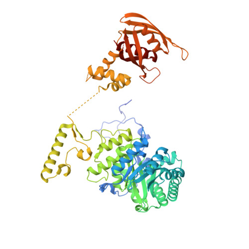
Crystal structure of LeuA from Mycobacterium tuberculosis, a key enzyme in leucine biosynthesis.
Koon, N., Squire, C.J., Baker, E.N.(2004) Proc Natl Acad Sci U S A 101: 8295-8300
- PubMed: 15159544
- DOI: https://doi.org/10.1073/pnas.0400820101
- Primary Citation of Related Structures:
1SR9, 3FIG - PubMed Abstract:
The leucine biosynthetic pathway is essential for the growth of Mycobacterium tuberculosis and is a potential target for the design of new anti-tuberculosis drugs. The crystal structure of alpha-isopropylmalate synthase, which catalyzes the first committed step in this pathway, has been determined by multiwavelength anomalous dispersion methods and refined at 2.0-A resolution in complex with its substrate alpha-ketoisovalerate. The structure reveals a tightly associated, domain-swapped dimer in which each monomer comprises an (alpha/beta)(8) TIM barrel catalytic domain, a helical linker domain, and a regulatory domain of novel fold. Mutational and crystallographic data indicate the latter as the site for leucine feedback inhibition of activity. Domain swapping enables the linker domain of one monomer to sit over the catalytic domain of the other, inserting residues into the active site that may be important in catalysis. The alpha-ketoisovalerate substrate binds to an active site zinc ion, adjacent to a cavity that can accommodate acetyl-CoA. Sequence and structural similarities point to a catalytic mechanism similar to that of malate synthase and an evolutionary relationship with an aldolase that catalyzes the reverse reaction on a similar substrate.
- Centre for Molecular Biodiscovery, School of Biological Sciences, and Department of Chemistry, University of Auckland, Auckland, New Zealand.
Organizational Affiliation:


