3FOE

Structural insight into the quinolone-DNA cleavage complex of type IIA topoisomerases


Experimental Data Snapshot

  • Method: X-RAY DIFFRACTION
  • Resolution: 4.00 Å
  • R-Value Free: 0.313 
  • R-Value Work: 0.258 
  • R-Value Observed: 0.264 

Starting Models: experimental
View more details

wwPDB Validation   3D Report Full Report


Ligand Structure Quality Assessment 


This is version 1.2 of the entry. See complete history


Literature

Structural insight into the quinolone-DNA cleavage complex of type IIA topoisomerases

Laponogov, I.Sohi, M.K.Veselkov, D.A.Pan, X.-S.Sawhney, R.Thompson, A.W.McAuley, K.E.Fisher, L.M.Sanderson, M.R.

(2009) Nat Struct Mol Biol 16: 667-669

  • DOI: https://doi.org/10.1038/nsmb.1604
  • Primary Citation of Related Structures:  
    3FOE, 3FOF

  • PubMed Abstract: 

    Type II topoisomerases alter DNA topology by forming a covalent DNA-cleavage complex that allows DNA transport through a double-stranded DNA break. We present the structures of cleavage complexes formed by the Streptococcus pneumoniae ParC breakage-reunion and ParE TOPRIM domains of topoisomerase IV stabilized by moxifloxacin and clinafloxacin, two antipneumococcal fluoroquinolones. These structures reveal two drug molecules intercalated at the highly bent DNA gate and help explain antibacterial quinolone action and resistance.


  • Organizational Affiliation

    Randall Division of Cell and Molecular Biophysics, King's College London, University of London, London, UK.


Macromolecules

Find similar proteins by:  (by identity cutoff)  |  3D Structure
Entity ID: 1
MoleculeChains Sequence LengthOrganismDetailsImage
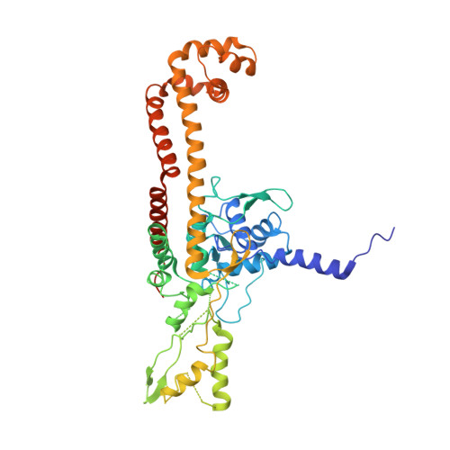
DNA topoisomerase 4 subunit A
A, B
496Streptococcus pneumoniaeMutation(s): 0 
Gene Names: parCSP_0855
EC: 5.99.1 (PDB Primary Data), 5.6.2.2 (UniProt)
UniProt
Find proteins for P72525 (Streptococcus pneumoniae serotype 4 (strain ATCC BAA-334 / TIGR4))
Explore P72525 
Go to UniProtKB:  P72525
Entity Groups  
Sequence Clusters30% Identity50% Identity70% Identity90% Identity95% Identity100% Identity
UniProt GroupP72525
Sequence Annotations
Expand
  • Reference Sequence
Find similar proteins by:  (by identity cutoff)  |  3D Structure
Entity ID: 2
MoleculeChains Sequence LengthOrganismDetailsImage
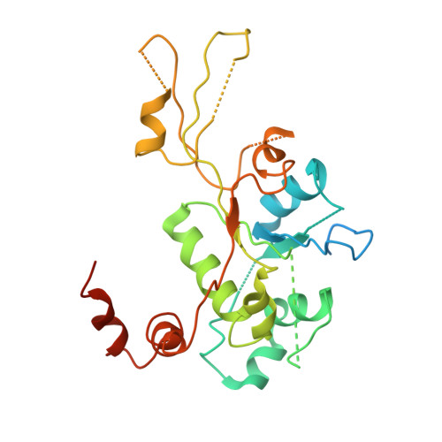
DNA topoisomerase 4 subunit B
C, D
268Streptococcus pneumoniaeMutation(s): 0 
Gene Names: parESP_0852
EC: 5.99.1 (PDB Primary Data), 5.6.2.2 (UniProt)
UniProt
Find proteins for Q59961 (Streptococcus pneumoniae serotype 4 (strain ATCC BAA-334 / TIGR4))
Explore Q59961 
Go to UniProtKB:  Q59961
Entity Groups  
Sequence Clusters30% Identity50% Identity70% Identity90% Identity95% Identity100% Identity
UniProt GroupQ59961
Sequence Annotations
Expand
  • Reference Sequence

Find similar nucleic acids by:  Sequence   |   3D Structure  

Entity ID: 3
MoleculeChains LengthOrganismImage
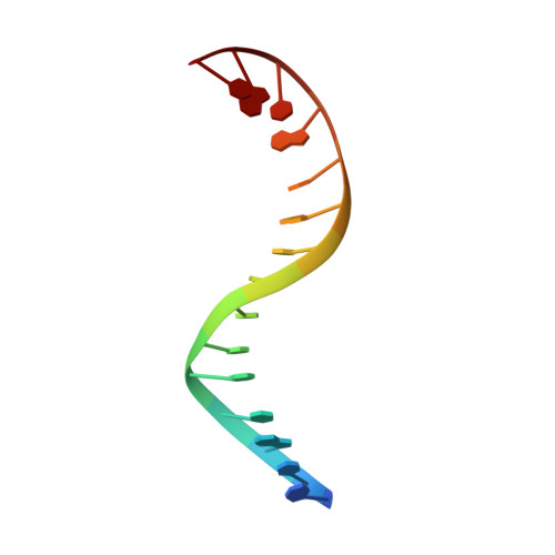
DNA (5'-D(P*AP*CP*CP*AP*AP*GP*GP*TP*CP*AP*TP*GP*AP*AP*T)-3')15N/A
Sequence Annotations
Expand
  • Reference Sequence

Find similar nucleic acids by:  Sequence   |   3D Structure  

Entity ID: 4
MoleculeChains LengthOrganismImage
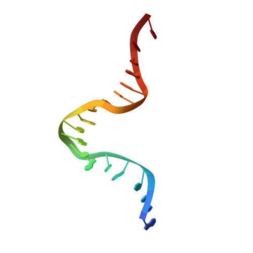
DNA (5'-D(P*AP*GP*TP*CP*AP*TP*TP*CP*AP*TP*GP*AP*CP*CP*TP*TP*GP*GP*T)-3')19N/A
Sequence Annotations
Expand
  • Reference Sequence

Find similar nucleic acids by:  Sequence   |   3D Structure  

Entity ID: 5
MoleculeChains LengthOrganismImage
DNA (5'-D(P*CP*TP*GP*TP*TP*TP*TP*AP*CP*GP*TP*GP*CP*AP*T)-3')15N/A
Sequence Annotations
Expand
  • Reference Sequence

Find similar nucleic acids by:  Sequence   |   3D Structure  

Entity ID: 6
MoleculeChains LengthOrganismImage
DNA (5'-D(P*GP*AP*CP*TP*AP*TP*GP*CP*AP*CP*GP*TP*AP*AP*AP*AP*CP*AP*G)-3')19N/A
Sequence Annotations
Expand
  • Reference Sequence
Small Molecules
Ligands 1 Unique
IDChains Name / Formula / InChI Key2D Diagram3D Interactions
NFX
Query on NFX

Download Ideal Coordinates CCD File 
I [auth F],
J [auth H]
7-[(3R)-3-aminopyrrolidin-1-yl]-8-chloro-1-cyclopropyl-6-fluoro-4-oxo-1,4-dihydroquinoline-3-carboxylic acid
C17 H17 Cl F N3 O3
QGPKADBNRMWEQR-MRVPVSSYSA-N
Experimental Data & Validation

Experimental Data

  • Method: X-RAY DIFFRACTION
  • Resolution: 4.00 Å
  • R-Value Free: 0.313 
  • R-Value Work: 0.258 
  • R-Value Observed: 0.264 
  • Space Group: P 32
Unit Cell:
Length ( Å )Angle ( ˚ )
a = 121.676α = 90
b = 121.676β = 90
c = 179.393γ = 120
Software Package:
Software NamePurpose
PHASERphasing
PHENIXrefinement
HKL-2000data reduction
HKL-2000data scaling

Structure Validation

View Full Validation Report


Ligand Structure Quality Assessment 


Entry History 

Deposition Data

Revision History  (Full details and data files)

  • Version 1.0: 2009-02-24
    Type: Initial release
  • Version 1.1: 2011-07-13
    Changes: Version format compliance
  • Version 1.2: 2023-11-01
    Changes: Data collection, Database references, Derived calculations, Refinement description