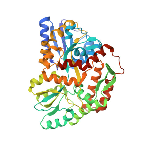
3G7V

Islet Amyloid Polypeptide (IAPP or Amylin) fused to Maltose Binding Protein


Experimental Data Snapshot

  • Method: X-RAY DIFFRACTION
  • Resolution: 1.86 Å
  • R-Value Free: 0.205 
  • R-Value Work: 0.176 
  • R-Value Observed: 0.178 

Starting Model: experimental
View more details

wwPDB Validation   3D Report Full Report


This is version 2.2 of the entry. See complete history


Literature

Atomic structures of IAPP (amylin) fusions suggest a mechanism for fibrillation and the role of insulin in the process

Wiltzius, J.J.Sievers, S.A.Sawaya, M.R.Eisenberg, D.

(2009) Protein Sci 18: 1521-1530

  • DOI: https://doi.org/10.1002/pro.145
  • Primary Citation of Related Structures:  
    3G7V, 3G7W

  • PubMed Abstract: 

    Islet Amyloid Polypeptide (IAPP or amylin) is a peptide hormone produced and stored in the beta-islet cells of the pancreas along with insulin. IAPP readily forms amyloid fibrils in vitro, and the deposition of fibrillar IAPP has been correlated with the pathology of type II diabetes. The mechanism of the conversion that IAPP undergoes from soluble to fibrillar forms has been unclear. By chaperoning IAPP through fusion to maltose binding protein, we find that IAPP can adopt a alpha-helical structure at residues 8-18 and 22-27 and that molecules of IAPP dimerize. Mutational analysis suggests that this dimerization is on the pathway to fibrillation. The structure suggests how IAPP may heterodimerize with insulin, which we confirmed by protein crosslinking. Taken together, these experiments suggest the helical dimerization of IAPP accelerates fibril formation and that insulin impedes fibrillation by blocking the IAPP dimerization interface.


  • Organizational Affiliation

    Howard Hughes Medical Institute, UCLA-DOE Institute of Genomics and Proteomics, Los Angeles, California 90095-1570, USA.


Macromolecules
Find similar proteins by:  (by identity cutoff)  |  3D Structure
Entity ID: 1
MoleculeChains Sequence LengthOrganismDetailsImage
Maltose-binding periplasmic protein, Islet amyloid polypeptide fusion protein
A, B, C, D
408Escherichia coli K-12Homo sapiens
This entity is chimeric
Mutation(s): 3 
Gene Names: malEb4034JW3994IAPP
UniProt & NIH Common Fund Data Resources
Find proteins for P0AEX9 (Escherichia coli (strain K12))
Explore P0AEX9 
Go to UniProtKB:  P0AEX9
Find proteins for P10997 (Homo sapiens)
Explore P10997 
Go to UniProtKB:  P10997
PHAROS:  P10997
GTEx:  ENSG00000121351 
Entity Groups  
Sequence Clusters30% Identity50% Identity70% Identity90% Identity95% Identity100% Identity
UniProt GroupsP0AEX9P10997
Sequence Annotations
Expand
  • Reference Sequence
Oligosaccharides

Help

Entity ID: 2
MoleculeChains Length2D Diagram Glycosylation3D Interactions
alpha-D-glucopyranose-(1-4)-alpha-D-glucopyranose
E, F, G, H
2N/A
Glycosylation Resources
GlyTouCan:  G07411ON
GlyCosmos:  G07411ON
Small Molecules
Ligands 2 Unique
IDChains Name / Formula / InChI Key2D Diagram3D Interactions
SO4
Query on SO4

Download Ideal Coordinates CCD File 
AA [auth D]
I [auth A]
J [auth A]
L [auth B]
M [auth B]
AA [auth D],
I [auth A],
J [auth A],
L [auth B],
M [auth B],
N [auth B],
O [auth B],
P [auth B],
S [auth C],
T [auth C],
U [auth C],
V [auth C],
W [auth C],
Y [auth D],
Z [auth D]
SULFATE ION
O4 S
QAOWNCQODCNURD-UHFFFAOYSA-L
GOL
Query on GOL

Download Ideal Coordinates CCD File 
BA [auth D],
K [auth A],
Q [auth B],
R [auth B],
X [auth C]
GLYCEROL
C3 H8 O3
PEDCQBHIVMGVHV-UHFFFAOYSA-N
Experimental Data & Validation

Experimental Data

  • Method: X-RAY DIFFRACTION
  • Resolution: 1.86 Å
  • R-Value Free: 0.205 
  • R-Value Work: 0.176 
  • R-Value Observed: 0.178 
  • Space Group: P 1 21 1
Unit Cell:
Length ( Å )Angle ( ˚ )
a = 104.025α = 90
b = 96.407β = 90.53
c = 103.996γ = 90
Software Package:
Software NamePurpose
DENZOdata reduction
SCALEPACKdata scaling
PHASERphasing
REFMACrefinement
PDB_EXTRACTdata extraction
BOSdata collection

Structure Validation

View Full Validation Report


Entry History 

Deposition Data

Revision History  (Full details and data files)

  • Version 1.0: 2009-06-23
    Type: Initial release
  • Version 1.1: 2011-07-13
    Changes: Advisory, Refinement description, Version format compliance
  • Version 1.2: 2017-08-16
    Changes: Refinement description, Source and taxonomy
  • Version 1.3: 2017-11-01
    Changes: Refinement description
  • Version 2.0: 2020-07-29
    Type: Remediation
    Reason: Carbohydrate remediation
    Changes: Atomic model, Data collection, Database references, Derived calculations, Non-polymer description, Structure summary
  • Version 2.1: 2021-10-20
    Changes: Database references, Structure summary
  • Version 2.2: 2023-09-06
    Changes: Data collection, Refinement description