3G9Y | pdb_00003g9y

Crystal structure of the second zinc finger from ZRANB2/ZNF265 bound to 6 nt ssRNA sequence AGGUAA


Experimental Data Snapshot

  • Method: X-RAY DIFFRACTION
  • Resolution: 1.40 Å
  • R-Value Free: 
    0.235 (Depositor), 0.234 (DCC) 
  • R-Value Work: 
    0.201 (Depositor), 0.207 (DCC) 
  • R-Value Observed: 
    0.203 (Depositor) 

wwPDB Validation 3D Report Full Report

Validation slider image for 3G9Y

This is version 1.3 of the entry. See complete history

Literature

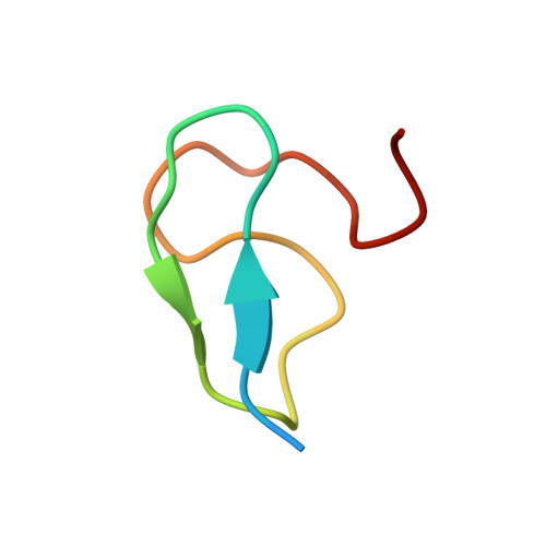
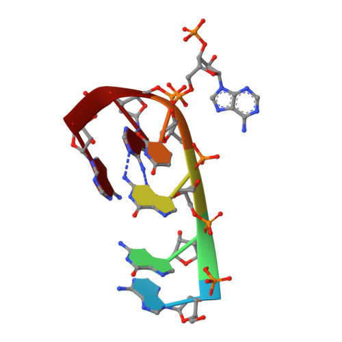
The zinc fingers of the SR-like protein ZRANB2 are single-stranded RNA-binding domains that recognize 5' splice site-like sequences

Loughlin, F.E.Mansfield, R.E.Vaz, P.M.McGrath, A.P.Setiyaputra, S.Gamsjaeger, R.Chen, E.S.Morris, B.J.Guss, J.M.Mackay, J.P.

(2009) Proc Natl Acad Sci U S A 106: 5581-5586

  • DOI: https://doi.org/10.1073/pnas.0802466106
  • Primary Citation Related Structures: 
    3G9Y

  • PubMed Abstract: 

    The alternative splicing of mRNA is a critical process in higher eukaryotes that generates substantial proteomic diversity. Many of the proteins that are essential to this process contain arginine/serine-rich (RS) domains. ZRANB2 is a widely-expressed and highly-conserved RS-domain protein that can regulate alternative splicing but lacks canonical RNA-binding domains. Instead, it contains 2 RanBP2-type zinc finger (ZnF) domains. We demonstrate that these ZnFs recognize ssRNA with high affinity and specificity. Each ZnF binds to a single AGGUAA motif and the 2 domains combine to recognize AGGUAA(N(x))AGGUAA double sites, suggesting that ZRANB2 regulates alternative splicing via a direct interaction with pre-mRNA at sites that resemble the consensus 5' splice site. We show using X-ray crystallography that recognition of an AGGUAA motif by a single ZnF is dominated by side-chain hydrogen bonds to the bases and formation of a guanine-tryptophan-guanine "ladder." A number of other human proteins that function in RNA processing also contain RanBP2 ZnFs in which the RNA-binding residues of ZRANB2 are conserved. The ZnFs of ZRANB2 therefore define another class of RNA-binding domain, advancing our understanding of RNA recognition and emphasizing the versatility of ZnF domains in molecular recognition.


  • Organizational Affiliation
    • School of Molecular and Microbial Biosciences, University of Sydney, Sydney NSW 2006, Australia.

Macromolecule Content 

  • Total Structure Weight: 5.76 kDa 
  • Atom Count: 426 
  • Modeled Residue Count: 35 
  • Deposited Residue Count: 39 
  • Unique protein chains: 1
  • Unique nucleic acid chains: 1

Macromolecules


Find similar proteins by:|  3D Structure
Entity ID: 1
MoleculeChains  Sequence LengthOrganismDetailsImage
Zinc finger Ran-binding domain-containing protein 233Homo sapiensMutation(s): 0 
Gene Names: ZRANB2
UniProt & NIH Common Fund Data Resources
Find proteins for O95218 (Homo sapiens)
Explore O95218 
Go to UniProtKB:  O95218
PHAROS:  O95218
GTEx:  ENSG00000132485 
Entity Groups
Sequence Clusters30% Identity50% Identity70% Identity90% Identity95% Identity100% Identity
UniProt GroupO95218
Sequence Annotations
Expand
Reference Sequence
Find similar nucleic acids by:  Sequence
Entity ID: 2
MoleculeChains LengthOrganismImage
RNA (5'-R(*AP*GP*GP*UP*AP*A)-3')B [auth C]6N/A
Sequence Annotations
Expand
Reference Sequence

Small Molecules

Ligands 1 Unique
IDChains Name / Formula / InChI Key2D Diagram3D Interactions
ZN

Query on ZN


Download:Ideal Coordinates CCD File
C [auth A]ZINC ION
Zn
PTFCDOFLOPIGGS-UHFFFAOYSA-N

Experimental Data & Validation

Experimental Data

  • Method: X-RAY DIFFRACTION
  • Resolution: 1.40 Å
  • R-Value Free:  0.235 (Depositor), 0.234 (DCC) 
  • R-Value Work:  0.201 (Depositor), 0.207 (DCC) 
  • R-Value Observed: 0.203 (Depositor) 
Space Group: P 65 2 2
Unit Cell:
Length ( Å )Angle ( ˚ )
a = 54.519α = 90
b = 54.519β = 90
c = 48.071γ = 120
Software Package:
Software NamePurpose
DENZOdata reduction
SCALEPACKdata scaling
REFMACrefinement
PDB_EXTRACTdata extraction
HKL-2000data collection
HKL-2000data reduction
HKL-2000data scaling
SHELXEmodel building
PHASERphasing

Structure Validation

View Full Validation Report


Entry History 

Deposition Data

Revision History  (Full details and data files)

  • Version 1.0: 2009-03-03
    Type: Initial release
  • Version 1.1: 2011-07-13
    Changes: Advisory, Version format compliance
  • Version 1.2: 2017-11-01
    Changes: Refinement description
  • Version 1.3: 2024-03-20
    Changes: Data collection, Database references, Derived calculations