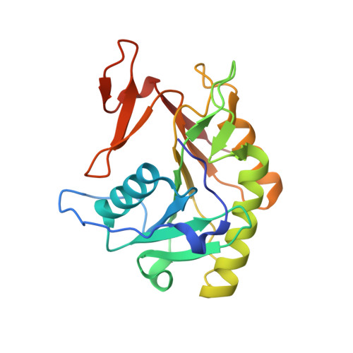
3GCD

Structure of the V. cholerae RTX cysteine protease domain in complex with an aza-Leucine peptide inhibitor


Experimental Data Snapshot

  • Method: X-RAY DIFFRACTION
  • Resolution: 2.35 Å
  • R-Value Free: 0.265 
  • R-Value Work: 0.218 
  • R-Value Observed: 0.221 

Starting Model: experimental
View more details

wwPDB Validation   3D Report Full Report


Ligand Structure Quality Assessment 


This is version 1.4 of the entry. See complete history


Literature

Mechanistic and structural insights into the proteolytic activation of Vibrio cholerae MARTX toxin.

Shen, A.Lupardus, P.J.Albrow, V.E.Guzzetta, A.Powers, J.C.Garcia, K.C.Bogyo, M.

(2009) Nat Chem Biol 5: 469-478

  • DOI: https://doi.org/10.1038/nchembio.178
  • Primary Citation of Related Structures:  
    3GCD

  • PubMed Abstract: 

    MARTX toxins modulate the virulence of a number of Gram-negative Vibrio species. This family of toxins is defined by the presence of a cysteine protease domain (CPD), which proteolytically activates the Vibrio cholerae MARTX toxin. Although recent structural studies of the CPD have uncovered a new allosteric activation mechanism, the mechanism of CPD substrate recognition or toxin processing is unknown. Here we show that interdomain cleavage of MARTXVc enhances effector domain function. We also identify the first small-molecule inhibitors of this protease domain and present the 2.35-A structure of the CPD bound to one of these inhibitors. This structure, coupled with biochemical and mutational studies of the toxin, reveals the molecular basis of CPD substrate specificity and underscores the evolutionary relationship between the CPD and the clan CD caspase proteases. These studies are likely to prove valuable for devising new antitoxin strategies for a number of bacterial pathogens.


  • Organizational Affiliation

    Department of Pathology, and Howard Hughes Medical Institute, Stanford School of Medicine, Stanford,California, USA.


Macromolecules
Find similar proteins by:  (by identity cutoff)  |  3D Structure
Entity ID: 1
MoleculeChains Sequence LengthOrganismDetailsImage
RTX toxin RtxA
A, B, C, D
209Vibrio choleraeMutation(s): 0 
Gene Names: rtxAvc1451VC_1451
EC: 3.4.22
UniProt
Find proteins for Q9KS12 (Vibrio cholerae serotype O1 (strain ATCC 39315 / El Tor Inaba N16961))
Explore Q9KS12 
Go to UniProtKB:  Q9KS12
Entity Groups  
Sequence Clusters30% Identity50% Identity70% Identity90% Identity95% Identity100% Identity
UniProt GroupQ9KS12
Sequence Annotations
Expand
  • Reference Sequence
Small Molecules
Ligands 3 Unique
IDChains Name / Formula / InChI Key2D Diagram3D Interactions
IHP
Query on IHP

Download Ideal Coordinates CCD File 
F [auth A],
I [auth B],
L [auth C],
O [auth D]
INOSITOL HEXAKISPHOSPHATE
C6 H18 O24 P6
IMQLKJBTEOYOSI-GPIVLXJGSA-N
AZ0
Query on AZ0

Download Ideal Coordinates CCD File 
E [auth A],
H [auth B],
K [auth C],
N [auth D]
ethyl (5S,8S,14S)-14-hydroxy-5,8,11-tris(2-methylpropyl)-3,6,9,12-tetraoxo-1-phenyl-2-oxa-4,7,10,11-tetraazapentadecan-15-oate
C30 H48 N4 O8
DDKYUZVIGXLULX-SDHOMARFSA-N
NA
Query on NA

Download Ideal Coordinates CCD File 
G [auth A],
J [auth B],
M [auth C],
P [auth D]
SODIUM ION
Na
FKNQFGJONOIPTF-UHFFFAOYSA-N
Experimental Data & Validation

Experimental Data

  • Method: X-RAY DIFFRACTION
  • Resolution: 2.35 Å
  • R-Value Free: 0.265 
  • R-Value Work: 0.218 
  • R-Value Observed: 0.221 
  • Space Group: P 21 21 21
Unit Cell:
Length ( Å )Angle ( ˚ )
a = 48.56α = 90
b = 65.854β = 90
c = 254.88γ = 90
Software Package:
Software NamePurpose
HKL-2000data collection
PHASERphasing
REFMACrefinement
HKL-2000data reduction
HKL-2000data scaling

Structure Validation

View Full Validation Report


Ligand Structure Quality Assessment 


Entry History 

Deposition Data

Revision History  (Full details and data files)

  • Version 1.0: 2009-05-26
    Type: Initial release
  • Version 1.1: 2011-07-13
    Changes: Atomic model, Database references, Derived calculations, Non-polymer description, Structure summary, Version format compliance
  • Version 1.2: 2012-12-12
    Changes: Other
  • Version 1.3: 2020-10-14
    Changes: Derived calculations, Structure summary
  • Version 1.4: 2023-09-06
    Changes: Data collection, Database references, Refinement description