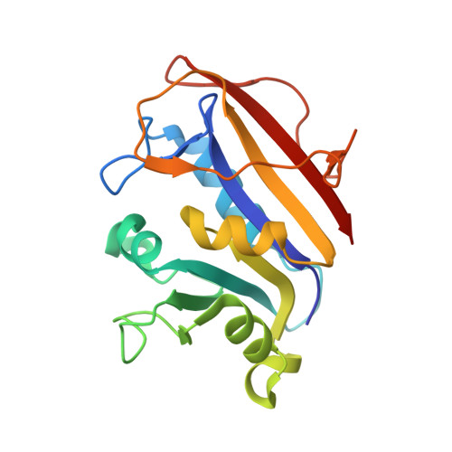
3GI2 | pdb_00003gi2

Human dihydrofolate reductase Q35K mutant inhibitor complex


Experimental Data Snapshot

  • Method: X-RAY DIFFRACTION
  • Resolution: 1.53 Å
  • R-Value Free: 
    0.203 (Depositor), 0.200 (DCC) 
  • R-Value Work: 
    0.157 (Depositor), 0.160 (DCC) 
  • R-Value Observed: 
    0.159 (Depositor) 

Starting Model: experimental
View more details

wwPDB Validation   3D Report Full Report


Ligand Structure Quality Assessment 

Created with Raphaël 2.3.0Worse 01 BetterLigand structure goodness of fit to experimental dataBest fitted NDPClick on this verticalbar to view detailsBest fitted GHWClick on this verticalbar to view details

This is version 1.3 of the entry. See complete history


Literature

Design, synthesis, and X-ray crystal structure of classical and nonclassical 2-amino-4-oxo-5-substituted-6-ethylthieno[2,3-d]pyrimidines as dual thymidylate synthase and dihydrofolate reductase inhibitors and as potential antitumor agents.

Gangjee, A.Li, W.Kisliuk, R.L.Cody, V.Pace, J.Piraino, J.Makin, J.

(2009) J Med Chem 52: 4892-4902

  • DOI: https://doi.org/10.1021/jm900490a
  • Primary Citation of Related Structures:  
    3GHC, 3GHV, 3GHW, 3GI2

  • PubMed Abstract: 

    N-{4-[(2-Amino-6-ethyl-4-oxo-3,4-dihydrothieno[2,3-d]pyrimidin-5-yl)thio]benzoyl}-L-glutamic acid 2 and 13 nonclassical analogues 2a-2m were synthesized as potential dual thymidylate synthase (TS) and dihydrofolate reductase (DHFR) inhibitors and as antitumor agents. The key intermediate in the synthesis was 2-amino-6-ethyl-5-iodothieno[2,3-d]pyrimidin-4(3H)-one, 7, to which various arylthiols were attached at the 5-position. Coupling 8 with L-glutamic acid diethyl ester and saponification afforded 2. X-ray crystal structures of 2 and 1 (the 6-methyl analogue of 2), DHFR, and NADPH showed for the first time that the thieno[2,3-d]pyrimidine ring binds in a "folate" mode. Compound 2 was an excellent dual inhibitor of human TS (IC50 = 54 nM) and human DHFR (IC50 = 19 nM) and afforded nanomolar GI50 values against tumor cells in culture. The 6-ethyl substitution in 2 increases both the potency (by 2-3 orders of magnitude) as well as the spectrum of tumor inhibition in vitro compared to the 6-methyl analogue 1. Some of the nonclassical analogues were potent and selective inhibitors of DHFR from Toxoplasma gondii.


  • Organizational Affiliation

    Division of Medicinal Chemistry, Graduate School of Pharmaceutical Sciences, Duquesne University, 600 Forbes Avenue, Pittsburgh, Pennsylvania 15282, USA. gangjee@duq.edu


Macromolecules
Find similar proteins by:  (by identity cutoff)  |  3D Structure
Entity ID: 1
MoleculeChains Sequence LengthOrganismDetailsImage
Dihydrofolate reductase187Homo sapiensMutation(s): 1 
Gene Names: DHFRDHFRP1
EC: 1.5.1.3
UniProt & NIH Common Fund Data Resources
Find proteins for P00374 (Homo sapiens)
Explore P00374 
Go to UniProtKB:  P00374
PHAROS:  P00374
GTEx:  ENSG00000228716 
Entity Groups  
Sequence Clusters30% Identity50% Identity70% Identity90% Identity95% Identity100% Identity
UniProt GroupP00374
Sequence Annotations
Expand
  • Reference Sequence
Small Molecules
Ligands 2 Unique
IDChains Name / Formula / InChI Key2D Diagram3D Interactions
NDP
Query on NDP

Download Ideal Coordinates CCD File 
C [auth A]NADPH DIHYDRO-NICOTINAMIDE-ADENINE-DINUCLEOTIDE PHOSPHATE
C21 H30 N7 O17 P3
ACFIXJIJDZMPPO-NNYOXOHSSA-N
GHW
Query on GHW

Download Ideal Coordinates CCD File 
B [auth A]N-({4-[(2-amino-6-methyl-4-oxo-3,4-dihydrothieno[2,3-d]pyrimidin-5-yl)sulfanyl]phenyl}carbonyl)-L-glutamic acid
C19 H18 N4 O6 S2
XZCPGLYOPFIZBL-NSHDSACASA-N
Experimental Data & Validation

Experimental Data

  • Method: X-RAY DIFFRACTION
  • Resolution: 1.53 Å
  • R-Value Free:  0.203 (Depositor), 0.200 (DCC) 
  • R-Value Work:  0.157 (Depositor), 0.160 (DCC) 
  • R-Value Observed: 0.159 (Depositor) 
Space Group: H 3
Unit Cell:
Length ( Å )Angle ( ˚ )
a = 84.574α = 90
b = 84.574β = 90
c = 77.943γ = 120
Software Package:
Software NamePurpose
HKL-2000data collection
MOLREPphasing
REFMACrefinement
MOSFLMdata reduction
SCALAdata scaling

Structure Validation

View Full Validation Report


Ligand Structure Quality Assessment 

Created with Raphaël 2.3.0Worse 01 BetterLigand structure goodness of fit to experimental dataBest fitted NDPClick on this verticalbar to view detailsBest fitted GHWClick on this verticalbar to view details

Entry History 

Deposition Data

  • Released Date: 2009-09-29 
  • Deposition Author(s): Cody, V.

Revision History  (Full details and data files)

  • Version 1.0: 2009-09-29
    Type: Initial release
  • Version 1.1: 2011-07-13
    Changes: Version format compliance
  • Version 1.2: 2021-10-20
    Changes: Database references, Derived calculations
  • Version 1.3: 2023-09-06
    Changes: Data collection, Refinement description