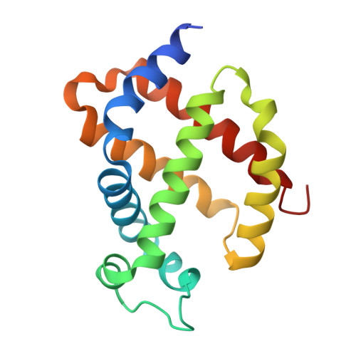
3GKT

Crystal structure of murine neuroglobin under Kr pressure


Experimental Data Snapshot

  • Method: X-RAY DIFFRACTION
  • Resolution: 1.86 Å
  • R-Value Free: 0.208 
  • R-Value Work: 0.173 
  • R-Value Observed: 0.175 

Starting Model: experimental
View more details

wwPDB Validation   3D Report Full Report


Ligand Structure Quality Assessment 


This is version 1.4 of the entry. See complete history


Literature

The structure of neuroglobin at high Xe and Kr pressure reveals partial conservation of globin internal cavities.

Moschetti, T.Mueller, U.Schulze, J.Brunori, M.Vallone, B.

(2009) Biophys J 97: 1700-1708

  • DOI: https://doi.org/10.1016/j.bpj.2009.05.059
  • Primary Citation of Related Structures:  
    3GK9, 3GKT, 3GLN

  • PubMed Abstract: 

    Neuroglobin (Ngb) is a hexacoordinate globin expressed in the brain of vertebrates. Ferrous Ngb binds dioxygen with high affinity and the O(2) adduct is able to scavenge NO. Convincing in vitro and in vivo data indicate that Ngb is involved in neuroprotection during hypoxia and ischemia. The 3D structure of Ngb reveals the presence of a wide internal cavity connecting its heme active site with the bulk. To explore the role of this "tunnel" in the control of ligand binding, we determined the structure of metNgb and NgbCO equilibrated with Xe or Kr. We show four docking sites for Xe (only two for Kr); two of the four Xe sites are within the large cavity. They are only partially conserved in globins, since the two proximal Xe sites identified in myoglobin (Xe1 and Xe2) are absent in Ngb, as well as in cytoglobin. The Xe docking sites in Ngb map a pathway within the protein matrix, leading to the heme, which becomes more accessible in the ligand-bound species. This may be of significance in connection with the redox chemistry that may be the primary function of this hexacoordinate globin.


  • Organizational Affiliation

    Department of Biochemical Sciences A.Rossi-Fanelli, University of Rome La Sapienza, Rome, Italy.


Macromolecules
Find similar proteins by:  (by identity cutoff)  |  3D Structure
Entity ID: 1
MoleculeChains Sequence LengthOrganismDetailsImage
Neuroglobin154Mus musculusMutation(s): 2 
Gene Names: Ngb
EC: 1.7
UniProt
Find proteins for Q9ER97 (Mus musculus)
Explore Q9ER97 
Go to UniProtKB:  Q9ER97
Entity Groups  
Sequence Clusters30% Identity50% Identity70% Identity90% Identity95% Identity100% Identity
UniProt GroupQ9ER97
Sequence Annotations
Expand
  • Reference Sequence
Experimental Data & Validation

Experimental Data

  • Method: X-RAY DIFFRACTION
  • Resolution: 1.86 Å
  • R-Value Free: 0.208 
  • R-Value Work: 0.173 
  • R-Value Observed: 0.175 
  • Space Group: H 3 2
Unit Cell:
Length ( Å )Angle ( ˚ )
a = 88.8α = 90
b = 88.8β = 90
c = 113.5γ = 120
Software Package:
Software NamePurpose
HKL-2000data collection
REFMACrefinement
XDSdata reduction
XSCALEdata scaling
REFMACphasing

Structure Validation

View Full Validation Report


Ligand Structure Quality Assessment 


Entry History 

Deposition Data

Revision History  (Full details and data files)

  • Version 1.0: 2009-09-22
    Type: Initial release
  • Version 1.1: 2011-07-13
    Changes: Advisory, Version format compliance
  • Version 1.2: 2018-10-17
    Changes: Data collection, Database references, Derived calculations
  • Version 1.3: 2021-11-10
    Changes: Database references, Derived calculations
  • Version 1.4: 2023-11-01
    Changes: Data collection, Refinement description