3GY5 | pdb_00003gy5

A comparative study on the inhibition of bovine beta-trypsin by bis-benzamidines diminazene and pentamidine by X-ray crystallography and ITC


Experimental Data Snapshot

  • Method: X-RAY DIFFRACTION
  • Resolution: 1.57 Å
  • R-Value Free: 
    0.197 (Depositor), 0.193 (DCC) 
  • R-Value Work: 
    0.163 (Depositor), 0.159 (DCC) 
  • R-Value Observed: 
    0.164 (Depositor) 

wwPDB Validation 3D Report Full Report

Validation slider image for 3GY5

Ligand Structure Quality Assessment 


This is version 1.2 of the entry. See complete history

Literature

Structural binding evidence of the trypanocidal drugs Berenil and Pentacarinate active principles to a serine protease model.

Perilo, C.S.Pereira, M.T.Santoro, M.M.Nagem, R.A.

(2010) Int J Biol Macromol 46: 502-511

  • DOI: https://doi.org/10.1016/j.ijbiomac.2010.03.006
  • Primary Citation Related Structures: 
    3GY2, 3GY3, 3GY4, 3GY5, 3GY6, 3GY7, 3GY8

  • PubMed Abstract: 

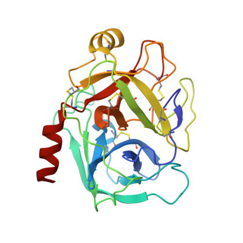
    Bovine trypsin is a model system for the serine protease class of enzymes, which is an important target for contemporary medicinal chemistry. Some structural and thermodynamic reports are available on its interaction with benzamidine-based compounds but no structural information is available so far on its binding modes to the active principles of the trypanocidal drugs Pentacarinate (pentamidine) and Berenil (diminazene). The crystallographic structures of bovine beta-trypsin in complex with the ligands were determined to a resolution of 1.57 A (diminazene) and 1.70 A (diminazene and pentamidine). The second benzamidine moieties in these inhibitors are bound to the enzyme in different hot spots and only few hydrogen bonds mediate these interactions. Thermodynamic parameters for the association of pentamidine with beta-trypsin reveal that this inhibitor has about 1.3-fold lower affinity than diminazene. Moreover its binding mode resembles other benzamidine-based compounds that assess the aryl binding pocket of the enzyme; however, with almost 2.5-fold higher affinity. This is the first structural evidence of the binding of Berenil and Pentacarinate active principles trypanocidal drugs to serine proteases.


  • Organizational Affiliation
    • Departamento de Bioquímica e Imunologia, Instituto de Ciências Biológicas, Universidade Federal de Minas Gerais, Avenida Antônio Carlos 6627, Belo Horizonte, MG, CEP 31270-901, Brazil.

Macromolecule Content 

  • Total Structure Weight: 24.66 kDa 
  • Atom Count: 2,109 
  • Modeled Residue Count: 223 
  • Deposited Residue Count: 223 
  • Unique protein chains: 1

Macromolecules

Find similar proteins by:|  3D Structure
Entity ID: 1
MoleculeChains  Sequence LengthOrganismDetailsImage
Cationic trypsin223Bos taurusMutation(s): 0 
EC: 3.4.21.4
UniProt
Find proteins for P00760 (Bos taurus)
Explore P00760 
Go to UniProtKB:  P00760
Entity Groups
Sequence Clusters30% Identity50% Identity70% Identity90% Identity95% Identity100% Identity
UniProt GroupP00760
Sequence Annotations
Expand
Reference Sequence

Small Molecules

Ligands 4 Unique
IDChains Name / Formula / InChI Key2D Diagram3D Interactions
BRN

Query on BRN


Download:Ideal Coordinates CCD File
C [auth A]BERENIL
C14 H15 N7
XNYZHCFCZNMTFY-UHFFFAOYSA-N
SO4

Query on SO4


Download:Ideal Coordinates CCD File
K [auth A]
L [auth A]
M [auth A]
N [auth A]
O [auth A]
K [auth A],
L [auth A],
M [auth A],
N [auth A],
O [auth A],
P [auth A]
SULFATE ION
O4 S
QAOWNCQODCNURD-UHFFFAOYSA-L
EDO

Query on EDO


Download:Ideal Coordinates CCD File
D [auth A]
E [auth A]
F [auth A]
G [auth A]
H [auth A]
D [auth A],
E [auth A],
F [auth A],
G [auth A],
H [auth A],
I [auth A],
J [auth A]
1,2-ETHANEDIOL
C2 H6 O2
LYCAIKOWRPUZTN-UHFFFAOYSA-N
CA

Query on CA


Download:Ideal Coordinates CCD File
B [auth A]CALCIUM ION
Ca
BHPQYMZQTOCNFJ-UHFFFAOYSA-N

Experimental Data & Validation

Experimental Data

  • Method: X-RAY DIFFRACTION
  • Resolution: 1.57 Å
  • R-Value Free:  0.197 (Depositor), 0.193 (DCC) 
  • R-Value Work:  0.163 (Depositor), 0.159 (DCC) 
  • R-Value Observed: 0.164 (Depositor) 
Space Group: P 21 21 21
Unit Cell:
Length ( Å )Angle ( ˚ )
a = 53.246α = 90
b = 56.976β = 90
c = 65.523γ = 90
Software Package:
Software NamePurpose
HKL-2000data collection
AMoREphasing
REFMACrefinement
HKL-2000data reduction
HKL-2000data scaling

Structure Validation

View Full Validation Report


Ligand Structure Quality Assessment 


Entry History 

Deposition Data

Revision History  (Full details and data files)

  • Version 1.0: 2010-03-23
    Type: Initial release
  • Version 1.1: 2011-07-13
    Changes: Version format compliance
  • Version 1.2: 2024-11-06
    Changes: Data collection, Database references, Derived calculations, Structure summary