3HQ9 | pdb_00003hq9

CcpA from G. sulfurreducens, S134P variant


Experimental Data Snapshot

  • Method: X-RAY DIFFRACTION
  • Resolution: 1.52 Å
  • R-Value Free: 
    0.184 (Depositor), 0.214 (DCC) 
  • R-Value Work: 
    0.160 (Depositor) 
  • R-Value Observed: 
    0.161 (Depositor) 

Starting Model: experimental
View more details

wwPDB Validation 3D Report Full Report

Validation slider image for 3HQ9

Ligand Structure Quality Assessment 


This is version 1.3 of the entry. See complete history

Literature

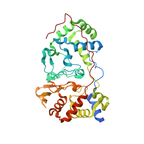
CcpA from Geobacter sulfurreducens is a basic di-heme cytochrome c peroxidase.

Hoffmann, M.Seidel, J.Einsle, O.

(2009) J Mol Biology 393: 951-965

  • DOI: https://doi.org/10.1016/j.jmb.2009.09.001
  • Primary Citation Related Structures: 
    3HQ6, 3HQ7, 3HQ8, 3HQ9

  • PubMed Abstract: 

    Bacterial di-heme cytochrome c peroxidases (CcpAs) protect the cell from reactive oxygen species by reducing hydrogen peroxide to water. The enzymes are c-type cytochromes, with both heme groups covalently attached to the protein chain via a characteristic binding motif. The genome of the dissimilatory metal-reducing bacterium Geobacter sulfurreducens revealed the presence of a ccpA gene and we isolated the gene product after recombinant expression in Escherichia coli. CcpA from G. sulfurreducens exhibited in vitro peroxidase activity with ABTS(2-) [2,2'-azino-bis(3-ethylbenzthiazoline-6-sulfonic acid)] as an electron donor, and the three-dimensional structure of the dimeric enzyme has been determined to high resolution. For activation, CcpA commonly requires reduction, with the exception of the Nitrosomonas europaea enzyme that retains its activity in the oxidized state. A G94K/K97Q/R100I triple point mutant was created to mimic the critical loop region of N. europaea CcpA, but its crystal structure revealed that the inactive, bis-histidinyl-coordinated form of the active-site heme group was retained. Subsequent mutational studies thus addressed an adjacent loop region, where a change in secondary structure accompanies the reductive activation of the enzyme. While an A124K/K128A double mutant did not show significant changes, the CcpA variants S134P/V135K and S134P led to a distortion of the loop region, accompanied by an opening of the active-site loop, leaving the enzyme in a constitutively active state.


  • Organizational Affiliation
    • Lehrstuhl für Biochemie, Institut für organische Chemie und Biochemie, Albert-Ludwigs-Universität Freiburg, Albertstrasse 21, Freiburg 79104, Germany.

Macromolecule Content 

  • Total Structure Weight: 77.24 kDa 
  • Atom Count: 5,448 
  • Modeled Residue Count: 618 
  • Deposited Residue Count: 690 
  • Unique protein chains: 1

Macromolecules

Find similar proteins by:|  3D Structure
Entity ID: 1
MoleculeChains  Sequence LengthOrganismDetailsImage
Cytochrome c551 peroxidase
A, B
345Geobacter sulfurreducensMutation(s): 1 
Gene Names: ccpAccpA-2GSU2813
EC: 1.11.1.5
UniProt
Find proteins for Q749D0 (Geobacter sulfurreducens (strain ATCC 51573 / DSM 12127 / PCA))
Explore Q749D0 
Go to UniProtKB:  Q749D0
Entity Groups
Sequence Clusters30% Identity50% Identity70% Identity90% Identity95% Identity100% Identity
UniProt GroupQ749D0
Sequence Annotations
Expand
Reference Sequence

Small Molecules

Ligands 5 Unique
IDChains Name / Formula / InChI Key2D Diagram3D Interactions
HEM

Query on HEM


Download:Ideal Coordinates CCD File
C [auth A],
D [auth A],
K [auth B],
L [auth B]
PROTOPORPHYRIN IX CONTAINING FE
C34 H32 Fe N4 O4
KABFMIBPWCXCRK-RGGAHWMASA-L
BU3

Query on BU3


Download:Ideal Coordinates CCD File
J [auth A],
R [auth B]
(R,R)-2,3-BUTANEDIOL
C4 H10 O2
OWBTYPJTUOEWEK-QWWZWVQMSA-N
OXL

Query on OXL


Download:Ideal Coordinates CCD File
H [auth A],
I [auth A],
P [auth B],
Q [auth B]
OXALATE ION
C2 O4
MUBZPKHOEPUJKR-UHFFFAOYSA-L
IMD

Query on IMD


Download:Ideal Coordinates CCD File
F [auth A],
G [auth A],
N [auth B],
O [auth B]
IMIDAZOLE
C3 H5 N2
RAXXELZNTBOGNW-UHFFFAOYSA-O
CA

Query on CA


Download:Ideal Coordinates CCD File
E [auth A],
M [auth B]
CALCIUM ION
Ca
BHPQYMZQTOCNFJ-UHFFFAOYSA-N

Experimental Data & Validation

Experimental Data

  • Method: X-RAY DIFFRACTION
  • Resolution: 1.52 Å
  • R-Value Free:  0.184 (Depositor), 0.214 (DCC) 
  • R-Value Work:  0.160 (Depositor) 
  • R-Value Observed: 0.161 (Depositor) 
Space Group: P 1
Unit Cell:
Length ( Å )Angle ( ˚ )
a = 50.899α = 104.71
b = 55.473β = 99.39
c = 68.606γ = 108.97
Software Package:
Software NamePurpose
REFMACrefinement

Structure Validation

View Full Validation Report


Ligand Structure Quality Assessment 


Entry History 

Deposition Data

Revision History  (Full details and data files)

  • Version 1.0: 2009-09-22
    Type: Initial release
  • Version 1.1: 2011-07-13
    Changes: Advisory, Refinement description, Version format compliance
  • Version 1.2: 2021-10-13
    Changes: Data collection, Database references, Derived calculations
  • Version 1.3: 2023-09-06
    Changes: Data collection, Refinement description