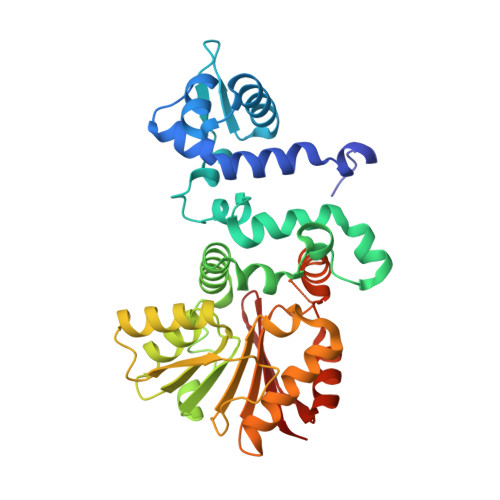
Molecular basis of substrate promiscuity for the SAM-dependent O-methyltransferase NcsB1, involved in the biosynthesis of the enediyne antitumor antibiotic neocarzinostatin.
Cooke, H.A., Guenther, E.L., Luo, Y., Shen, B., Bruner, S.D.(2009) Biochemistry 48: 9590-9598
- PubMed: 19702337
- DOI: https://doi.org/10.1021/bi901257q
- Primary Citation of Related Structures:
3I53, 3I58, 3I5U, 3I64 - PubMed Abstract:
The small molecule component of chromoprotein enediyne antitumor antibiotics is biosynthesized through a convergent route, incorporating amino acid, polyketide, and carbohydrate building blocks around a central enediyne hydrocarbon core. The naphthoic acid moiety of the enediyne neocarzinostatin plays key roles in the biological activity of the natural product by interacting with both the carrier protein and duplex DNA at the site of action. We have previously described the in vitro characterization of an S-adenosylmethionine-dependent O-methyltransferase (NcsB1) in the neocarzinostatin biosynthetic pathway [Luo, Y., Lin, S., Zhang, J., Cooke, H. A., Bruner, S. D., and Shen, B. (2008) J. Biol. Chem. 283, 14694-14702]. Here we provide a structural basis for NcsB1 activity, illustrating that the enzyme shares an overall architecture with a large family of S-adenosylmethionine-dependent proteins. In addition, NcsB1 represents the first enzyme to be structurally characterized in the biosynthetic pathway of neocarzinostatin. By cocrystallizing the enzyme with various combinations of the cofactor and substrate analogues, details of the active site structure have been established. Changes in subdomain orientation were observed via comparison of structures in the presence and absence of substrate, suggesting that reorientation of the enzyme is involved in binding of the substrate. In addition, residues important for substrate discrimination were predicted and probed through site-directed mutagenesis and in vitro biochemical characterization.
- Department of Chemistry, Merkert Chemistry Center, Boston College, Chestnut Hill, Massachusetts 02467, USA.
Organizational Affiliation:


