3I7R | pdb_00003i7r

Dihydrodipicolinate synthase - K161R


Experimental Data Snapshot

  • Method: X-RAY DIFFRACTION
  • Resolution: 2.10 Å
  • R-Value Free: 
    0.230 (Depositor), 0.236 (DCC) 
  • R-Value Work: 
    0.195 (Depositor), 0.200 (DCC) 
  • R-Value Observed: 
    0.197 (Depositor) 

Starting Model: experimental
View more details

wwPDB Validation 3D Report Full Report

Validation slider image for 3I7R

This is version 1.3 of the entry. See complete history

Literature

How essential is the 'essential' active-site lysine in dihydrodipicolinate synthase?

Soares da Costa, T.P.Muscroft-Taylor, A.C.Dobson, R.C.Devenish, S.R.Jameson, G.B.Gerrard, J.A.

(2010) Biochimie 92: 837-845

  • DOI: https://doi.org/10.1016/j.biochi.2010.03.004
  • Primary Citation Related Structures: 
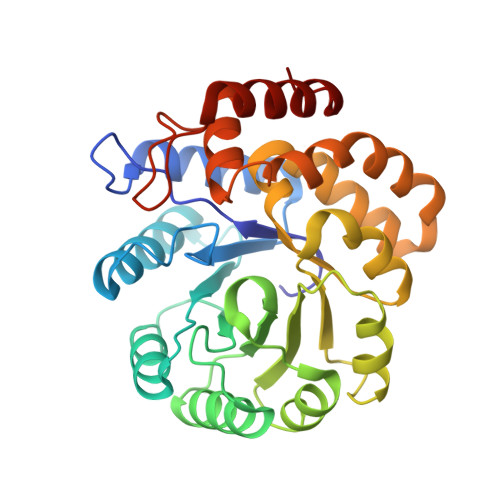
    3I7Q, 3I7R, 3I7S

  • PubMed Abstract: 

    Dihydrodipicolinate synthase (DHDPS, E.C. 4.2.1.52), a validated antibiotic target, catalyses the first committed step in the lysine biosynthetic pathway: the condensation reaction between (S)-aspartate beta-semialdehyde [(S)-ASA] and pyruvate via the formation of a Schiff base intermediate between pyruvate and the absolutely conserved active-site lysine. Escherichia coli DHDPS mutants K161A and K161R of the active-site lysine were characterised for the first time. Unexpectedly, the mutant enzymes were still catalytically active, albeit with a significant decrease in activity. The k(cat) values for DHDPS-K161A and DHDPS-K161R were 0.06 +/- 0.02 s(-1) and 0.16 +/- 0.06 s(-1) respectively, compared to 45 +/- 3 s(-1) for the wild-type enzyme. Remarkably, the K(M) values for pyruvate increased by only 3-fold for DHDPS-K161A and DHDPS-K161R (0.45 +/- 0.04 mM and 0.57 +/- 0.06 mM, compared to 0.15 +/- 0.01 mM for the wild-type DHDPS), while the K(M) values for (S)-ASA remained the same for DHDPS-K161R (0.12 +/- 0.01 mM) and increased by only 2-fold for DHDPS-K161A (0.23 +/- 0.02 mM) and the K(i) for lysine was unchanged. The X-ray crystal structures of DHDPS-K161A and DHDPS-K161R were solved at resolutions of 2.0 and 2.1 A respectively and showed no changes in their secondary or tertiary structures when compared to the wild-type structure. The crystal structure of DHDPS-K161A with pyruvate bound at the active site was solved at a resolution of 2.3 A and revealed a defined binding pocket for pyruvate that is thus not dependent upon lysine 161. Taken together with ITC and NMR data, it is concluded that although lysine 161 is important in the wild-type DHDPS-catalysed reaction, it is not absolutely essential for catalysis.


  • Organizational Affiliation
    • Biomolecular Interaction Centre and School of Biological Sciences, University of Canterbury, Christchurch, New Zealand.

Macromolecule Content 

  • Total Structure Weight: 63.54 kDa 
  • Atom Count: 4,900 
  • Modeled Residue Count: 584 
  • Deposited Residue Count: 584 
  • Unique protein chains: 1

Macromolecules

Find similar proteins by:|  3D Structure
Entity ID: 1
MoleculeChains  Sequence LengthOrganismDetailsImage
Dihydrodipicolinate synthase
A, B
292Escherichia coli K-12Mutation(s): 1 
Gene Names: b2478dapAJW2463
EC: 4.2.1.52 (PDB Primary Data), 4.3.3.7 (UniProt)
UniProt
Find proteins for P0A6L2 (Escherichia coli (strain K12))
Explore P0A6L2 
Go to UniProtKB:  P0A6L2
Entity Groups
Sequence Clusters30% Identity50% Identity70% Identity90% Identity95% Identity100% Identity
UniProt GroupP0A6L2
Sequence Annotations
Expand
Reference Sequence

Small Molecules

Ligands 4 Unique
IDChains Name / Formula / InChI Key2D Diagram3D Interactions
PO4

Query on PO4


Download:Ideal Coordinates CCD File
J [auth A]PHOSPHATE ION
O4 P
NBIIXXVUZAFLBC-UHFFFAOYSA-K
GOL

Query on GOL


Download:Ideal Coordinates CCD File
E [auth A]
F [auth A]
G [auth A]
H [auth A]
M [auth B]
E [auth A],
F [auth A],
G [auth A],
H [auth A],
M [auth B],
N [auth B]
GLYCEROL
C3 H8 O3
PEDCQBHIVMGVHV-UHFFFAOYSA-N
K

Query on K


Download:Ideal Coordinates CCD File
C [auth A],
D [auth A],
K [auth B],
L [auth B]
POTASSIUM ION
K
NPYPAHLBTDXSSS-UHFFFAOYSA-N
CL

Query on CL


Download:Ideal Coordinates CCD File
I [auth A],
O [auth B]
CHLORIDE ION
Cl
VEXZGXHMUGYJMC-UHFFFAOYSA-M

Experimental Data & Validation

Experimental Data

  • Method: X-RAY DIFFRACTION
  • Resolution: 2.10 Å
  • R-Value Free:  0.230 (Depositor), 0.236 (DCC) 
  • R-Value Work:  0.195 (Depositor), 0.200 (DCC) 
  • R-Value Observed: 0.197 (Depositor) 
Space Group: P 31 2 1
Unit Cell:
Length ( Å )Angle ( ˚ )
a = 121.435α = 90
b = 121.435β = 90
c = 111.037γ = 120
Software Package:
Software NamePurpose
CrystalCleardata collection
AMoREphasing
REFMACrefinement
d*TREKdata reduction
d*TREKdata scaling

Structure Validation

View Full Validation Report


Entry History 

Deposition Data

Revision History  (Full details and data files)

  • Version 1.0: 2010-04-14
    Type: Initial release
  • Version 1.1: 2011-07-13
    Changes: Advisory, Version format compliance
  • Version 1.2: 2021-10-13
    Changes: Database references, Derived calculations
  • Version 1.3: 2023-09-06
    Changes: Data collection, Refinement description