3ID5

Crystal structure of Sulfolobus solfataricus C/D RNP assembled with Nop5, fibrillarin, L7Ae and a split half C/D RNA


Experimental Data Snapshot

  • Method: X-RAY DIFFRACTION
  • Resolution: 4.01 Å
  • R-Value Free: 
    0.304 (Depositor), 0.280 (DCC) 
  • R-Value Work: 
    0.285 (Depositor), 0.270 (DCC) 
  • R-Value Observed: 
    0.286 (Depositor) 

wwPDB Validation   3D Report Full Report


Ligand Structure Quality Assessment 

Created with Raphaël 2.3.0Worse 01 BetterLigand structure goodness of fit to experimental dataBest fitted SAMClick on this verticalbar to view details

This is version 1.4 of the entry. See complete history


Literature

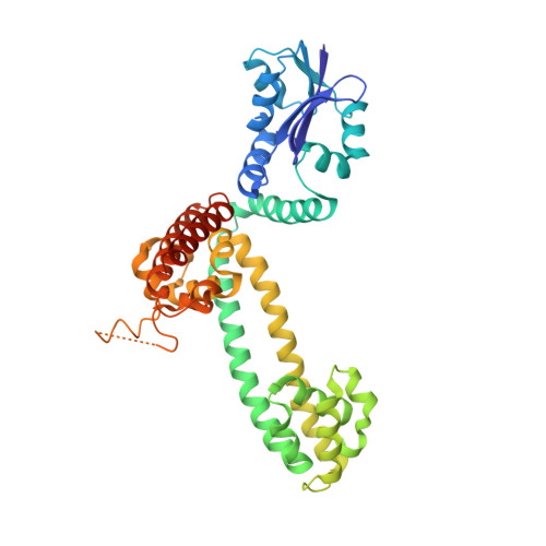
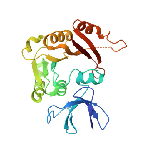
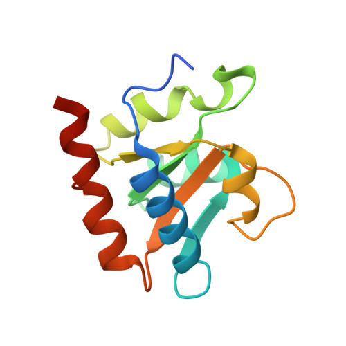
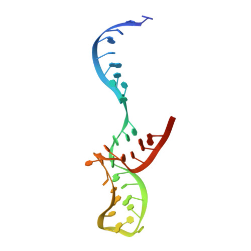
Structural organization of box C/D RNA-guided RNA methyltransferase.

Ye, K.Jia, R.Lin, J.Ju, M.Peng, J.Xu, A.Zhang, L.

(2009) Proc Natl Acad Sci U S A 106: 13808-13813

  • DOI: https://doi.org/10.1073/pnas.0905128106
  • Primary Citation of Related Structures:  
    3ICX, 3ID5, 3ID6

  • PubMed Abstract: 

    Box C/D guide RNAs are abundant noncoding RNAs that primarily function to direct the 2'-O-methylation of specific nucleotides by base-pairing with substrate RNAs. In archaea, a bipartite C/D RNA assembles with L7Ae, Nop5, and the methyltransferase fibrillarin into a modification enzyme with unique substrate specificity. Here, we determined the crystal structure of an archaeal C/D RNA-protein complex (RNP) composed of all 3 core proteins and an engineered half-guide RNA at 4 A resolution, as well as 2 protein substructures at higher resolution. The RNP structure reveals that the C-terminal domains of Nop5 in the dimeric complex provide symmetric anchoring sites for 2 L7Ae-associated kink-turn motifs of the C/D RNA. A prominent protrusion in Nop5 seems to be important for guide RNA organization and function and for discriminating the structurally related U4 snRNA. Multiple conformations of the N-terminal domain of Nop5 and its associated fibrillarin in different structures indicate the inherent flexibility of the catalytic module, suggesting that a swinging motion of the catalytic module is part of the enzyme mechanism. We also built a model of a native C/D RNP with substrate and fibrillarin in an active conformation. Our results provide insight into the overall organization and mechanism of action of C/D RNA-guided RNA methyltransferases.


  • Organizational Affiliation

    National Institute of Biological Sciences, Beijing 102206, China.


Macromolecules

Find similar proteins by:  (by identity cutoff)  |  3D Structure
Entity ID: 1
MoleculeChains Sequence LengthOrganismDetailsImage
Pre mRNA splicing protein
A, E
388Saccharolobus solfataricusMutation(s): 1 
Gene Names: SSO0939
UniProt
Find proteins for Q97ZH3 (Saccharolobus solfataricus (strain ATCC 35092 / DSM 1617 / JCM 11322 / P2))
Explore Q97ZH3 
Go to UniProtKB:  Q97ZH3
Entity Groups  
Sequence Clusters30% Identity50% Identity70% Identity90% Identity95% Identity100% Identity
UniProt GroupQ97ZH3
Sequence Annotations
Expand
  • Reference Sequence
Find similar proteins by:  (by identity cutoff)  |  3D Structure
Entity ID: 2
MoleculeChains Sequence LengthOrganismDetailsImage
Fibrillarin-like rRNA/tRNA 2'-O-methyltransferase
B, F
232Saccharolobus solfataricusMutation(s): 1 
Gene Names: flpASSO0940C33_014
EC: 2.1.1
UniProt
Find proteins for P58032 (Saccharolobus solfataricus (strain ATCC 35092 / DSM 1617 / JCM 11322 / P2))
Explore P58032 
Go to UniProtKB:  P58032
Entity Groups  
Sequence Clusters30% Identity50% Identity70% Identity90% Identity95% Identity100% Identity
UniProt GroupP58032
Sequence Annotations
Expand
  • Reference Sequence
Find similar proteins by:  (by identity cutoff)  |  3D Structure
Entity ID: 3
MoleculeChains Sequence LengthOrganismDetailsImage
50S ribosomal protein L7Ae
C, G
130Saccharolobus solfataricusMutation(s): 0 
Gene Names: rpl7aeSSO0091C04_031
UniProt
Find proteins for P55858 (Saccharolobus solfataricus (strain ATCC 35092 / DSM 1617 / JCM 11322 / P2))
Explore P55858 
Go to UniProtKB:  P55858
Entity Groups  
Sequence Clusters30% Identity50% Identity70% Identity90% Identity95% Identity100% Identity
UniProt GroupP55858
Sequence Annotations
Expand
  • Reference Sequence
Experimental Data & Validation

Experimental Data

  • Method: X-RAY DIFFRACTION
  • Resolution: 4.01 Å
  • R-Value Free:  0.304 (Depositor), 0.280 (DCC) 
  • R-Value Work:  0.285 (Depositor), 0.270 (DCC) 
  • R-Value Observed: 0.286 (Depositor) 
Space Group: P 41 21 2
Unit Cell:
Length ( Å )Angle ( ˚ )
a = 265.485α = 90
b = 265.485β = 90
c = 129.434γ = 90
Software Package:
Software NamePurpose
REFMACrefinement
PDB_EXTRACTdata extraction
HKL-2000data reduction
HKL-2000data scaling
SHARPphasing

Structure Validation

View Full Validation Report


Ligand Structure Quality Assessment 

Created with Raphaël 2.3.0Worse 01 BetterLigand structure goodness of fit to experimental dataBest fitted SAMClick on this verticalbar to view details

Entry History 

Deposition Data

  • Released Date: 2009-08-25 
  • Deposition Author(s): Ye, K.

Revision History  (Full details and data files)

  • Version 1.0: 2009-08-25
    Type: Initial release
  • Version 1.1: 2011-07-13
    Changes: Advisory, Version format compliance
  • Version 1.2: 2013-11-20
    Changes: Database references
  • Version 1.3: 2021-11-10
    Changes: Database references, Derived calculations
  • Version 1.4: 2024-05-29
    Changes: Data collection, Refinement description