3K0F | pdb_00003k0f

Crystal structure of the phosphorylation-site double mutant T426A/T432A of the KaiC circadian clock protein


Experimental Data Snapshot

  • Method: X-RAY DIFFRACTION
  • Resolution: 3.00 Å
  • R-Value Free: 
    0.288 (Depositor), 0.292 (DCC) 
  • R-Value Work: 
    0.229 (Depositor), 0.238 (DCC) 
  • R-Value Observed: 
    0.229 (Depositor) 

Starting Model: experimental
View more details

wwPDB Validation 3D Report Full Report

Validation slider image for 3K0F

Ligand Structure Quality Assessment 


This is version 1.4 of the entry. See complete history

Literature

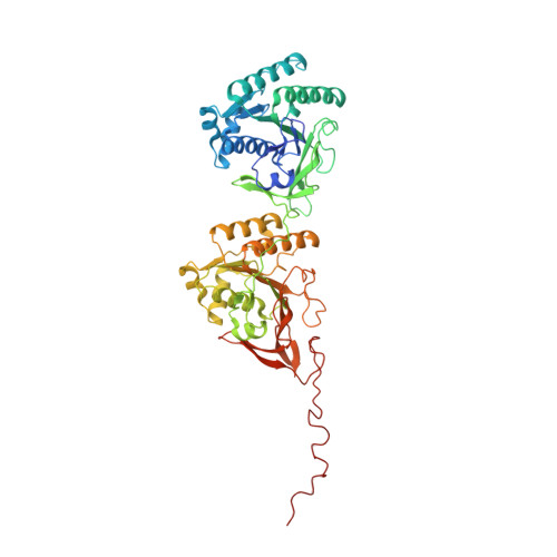
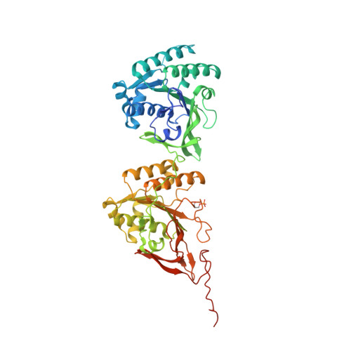
Structures of KaiC Circadian Clock Mutant Proteins: A New Phosphorylation Site at T426 and Mechanisms of Kinase, ATPase and Phosphatase.

Pattanayek, R.Mori, T.Xu, Y.Pattanayek, S.Johnson, C.H.Egli, M.

(2009) PLoS One 4: e7529-e7529

  • DOI: https://doi.org/10.1371/journal.pone.0007529
  • Primary Citation Related Structures: 
    3JZM, 3K09, 3K0A, 3K0C, 3K0E, 3K0F

  • PubMed Abstract: 

    The circadian clock of the cyanobacterium Synechococcus elongatus can be reconstituted in vitro by three proteins, KaiA, KaiB and KaiC. Homo-hexameric KaiC displays kinase, phosphatase and ATPase activities; KaiA enhances KaiC phosphorylation and KaiB antagonizes KaiA. Phosphorylation and dephosphorylation of the two known sites in the C-terminal half of KaiC subunits, T432 and S431, follow a strict order (TS-->pTS-->pTpS-->TpS-->TS) over the daily cycle, the origin of which is not understood. To address this void and to analyze the roles of KaiC active site residues, in particular T426, we determined structures of single and double P-site mutants of S. elongatus KaiC. The conformations of the loop region harboring P-site residues T432 and S431 in the crystal structures of six KaiC mutant proteins exhibit subtle differences that result in various distances between Thr (or Ala/Asn/Glu) and Ser (or Ala/Asp) residues and the ATP gamma-phosphate. T432 is phosphorylated first because it lies consistently closer to Pgamma. The structures of the S431A and T432E/S431A mutants reveal phosphorylation at T426. The environments of the latter residue in the structures and functional data for T426 mutants in vitro and in vivo imply a role in dephosphorylation. We provide evidence for a third phosphorylation site in KaiC at T426. T426 and S431 are closely spaced and a KaiC subunit cannot carry phosphates at both sites simultaneously. Fewer subunits are phosphorylated at T426 in the two KaiC mutants compared to phosphorylated T432 and/or S431 residues in the structures of wt and other mutant KaiCs, suggesting that T426 phosphorylation may be labile. The structures combined with functional data for a host of KaiC mutant proteins help rationalize why S431 trails T432 in the loss of its phosphate and shed light on the mechanisms of the KaiC kinase, ATPase and phosphatase activities.


  • Organizational Affiliation
    • Department of Biochemistry, Vanderbilt University, School of Medicine, Nashville, Tennessee, United States of America.

Macromolecule Content 

  • Total Structure Weight: 354.78 kDa 
  • Atom Count: 23,930 
  • Modeled Residue Count: 2,968 
  • Deposited Residue Count: 3,114 
  • Unique protein chains: 2

Macromolecules

Find similar proteins by:|  3D Structure
Entity ID: 1
MoleculeChains  Sequence LengthOrganismDetailsImage
Circadian clock protein kinase KaiC519Synechococcus elongatus PCC 7942 = FACHB-805Mutation(s): 2 
Gene Names: kaicsee0011Synpcc7942_1216
EC: 2.7.11.17 (PDB Primary Data), 3.6.4 (UniProt), 2.7.11.1 (UniProt)
UniProt
Find proteins for Q79PF4 (Synechococcus elongatus (strain ATCC 33912 / PCC 7942 / FACHB-805))
Explore Q79PF4 
Go to UniProtKB:  Q79PF4
Entity Groups
Sequence Clusters30% Identity50% Identity70% Identity90% Identity95% Identity100% Identity
UniProt GroupQ79PF4
Sequence Annotations
Expand
Reference Sequence
Find similar proteins by:|  3D Structure
Entity ID: 2
MoleculeChains  Sequence LengthOrganismDetailsImage
Circadian clock protein kinase KaiC
B, C, D, E, F
519Synechococcus elongatus PCC 7942 = FACHB-805Mutation(s): 2 
Gene Names: kaicsee0011Synpcc7942_1216
EC: 2.7.11.17 (PDB Primary Data), 3.6.4 (UniProt), 2.7.11.1 (UniProt)
UniProt
Find proteins for Q79PF4 (Synechococcus elongatus (strain ATCC 33912 / PCC 7942 / FACHB-805))
Explore Q79PF4 
Go to UniProtKB:  Q79PF4
Entity Groups
Sequence Clusters30% Identity50% Identity70% Identity90% Identity95% Identity100% Identity
UniProt GroupQ79PF4
Sequence Annotations
Expand
Reference Sequence

Small Molecules

Ligands 2 Unique
IDChains Name / Formula / InChI Key2D Diagram3D Interactions
ATP

Query on ATP


Download:Ideal Coordinates CCD File
AA [auth F]
H [auth A]
I [auth A]
L [auth B]
M [auth B]
AA [auth F],
H [auth A],
I [auth A],
L [auth B],
M [auth B],
P [auth C],
Q [auth C],
T [auth D],
U [auth D],
W [auth E],
X [auth E],
Z [auth F]
ADENOSINE-5'-TRIPHOSPHATE
C10 H16 N5 O13 P3
ZKHQWZAMYRWXGA-KQYNXXCUSA-N
MG

Query on MG


Download:Ideal Coordinates CCD File
G [auth A]
J [auth A]
K [auth B]
N [auth B]
O [auth C]
G [auth A],
J [auth A],
K [auth B],
N [auth B],
O [auth C],
R [auth C],
S [auth D],
V [auth E],
Y [auth F]
MAGNESIUM ION
Mg
JLVVSXFLKOJNIY-UHFFFAOYSA-N
Modified Residues  1 Unique
IDChains TypeFormula2D DiagramParent
SEP
Query on SEP
B, C, D, E, F
L-PEPTIDE LINKINGC3 H8 N O6 PSER

Experimental Data & Validation

Experimental Data

  • Method: X-RAY DIFFRACTION
  • Resolution: 3.00 Å
  • R-Value Free:  0.288 (Depositor), 0.292 (DCC) 
  • R-Value Work:  0.229 (Depositor), 0.238 (DCC) 
  • R-Value Observed: 0.229 (Depositor) 
Space Group: P 21 21 21
Unit Cell:
Length ( Å )Angle ( ˚ )
a = 132.927α = 90
b = 135.415β = 90
c = 205.618γ = 90
Software Package:
Software NamePurpose
HKL-2000data collection
CNSrefinement
HKL-2000data reduction
SCALEPACKdata scaling
CNSphasing

Structure Validation

View Full Validation Report


Ligand Structure Quality Assessment 


Entry History 

Deposition Data

Revision History  (Full details and data files)

  • Version 1.0: 2010-03-31
    Type: Initial release
  • Version 1.1: 2011-07-13
    Changes: Version format compliance
  • Version 1.2: 2021-10-13
    Changes: Database references, Derived calculations
  • Version 1.3: 2023-09-06
    Changes: Data collection, Refinement description
  • Version 1.4: 2024-11-20
    Changes: Structure summary