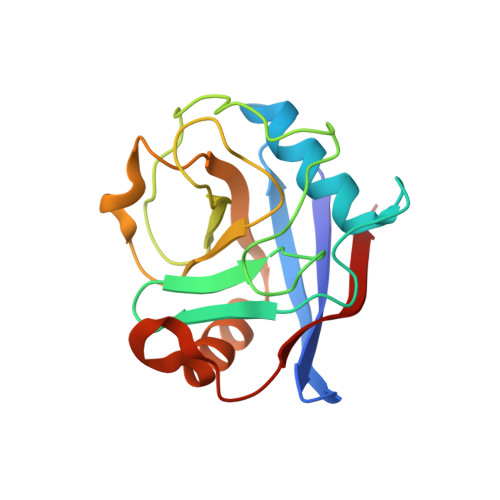
Hidden alternative structures of proline isomerase essential for catalysis.
Fraser, J.S., Clarkson, M.W., Degnan, S.C., Erion, R., Kern, D., Alber, T.(2009) Nature 462: 669-673
- PubMed: 19956261
- DOI: https://doi.org/10.1038/nature08615
- Primary Citation of Related Structures:
3K0M, 3K0N, 3K0O, 3K0P, 3K0Q, 3K0R - PubMed Abstract:
A long-standing challenge is to understand at the atomic level how protein dynamics contribute to enzyme catalysis. X-ray crystallography can provide snapshots of conformational substates sampled during enzymatic reactions, while NMR relaxation methods reveal the rates of interconversion between substates and the corresponding relative populations. However, these current methods cannot simultaneously reveal the detailed atomic structures of the rare states and rationalize the finding that intrinsic motions in the free enzyme occur on a timescale similar to the catalytic turnover rate. Here we introduce dual strategies of ambient-temperature X-ray crystallographic data collection and automated electron-density sampling to structurally unravel interconverting substates of the human proline isomerase, cyclophilin A (CYPA, also known as PPIA). A conservative mutation outside the active site was designed to stabilize features of the previously hidden minor conformation. This mutation not only inverts the equilibrium between the substates, but also causes large, parallel reductions in the conformational interconversion rates and the catalytic rate. These studies introduce crystallographic approaches to define functional minor protein conformations and, in combination with NMR analysis of the enzyme dynamics in solution, show how collective motions directly contribute to the catalytic power of an enzyme.
- Department of Molecular and Cell Biology/QB3, University of California, Berkeley, California 94720-3220, USA.
Organizational Affiliation:


