3K5T | pdb_00003k5t

Crystal structure of human diamine oxidase in space group C2221


Experimental Data Snapshot

  • Method: X-RAY DIFFRACTION
  • Resolution: 2.11 Å
  • R-Value Free: 
    0.290 (Depositor), 0.291 (DCC) 
  • R-Value Work: 
    0.236 (Depositor), 0.237 (DCC) 
  • R-Value Observed: 
    0.239 (Depositor) 

Starting Model: experimental
View more details

wwPDB Validation 3D Report Full Report

Validation slider image for 3K5T

Ligand Structure Quality Assessment 


This is version 2.2 of the entry. See complete history

Literature

A new crystal form of human diamine oxidase.

McGrath, A.P.Hilmer, K.M.Collyer, C.A.Dooley, D.M.Guss, J.M.

(2010) Acta Crystallogr Sect F Struct Biol Cryst Commun 66: 137-142

  • DOI: https://doi.org/10.1107/S1744309109052130
  • Primary Citation Related Structures: 
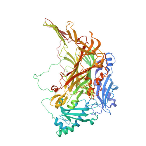
    3K5T

  • PubMed Abstract: 

    Copper amine oxidases (CAOs) are ubiquitous in nature and catalyse the oxidative deamination of primary amines to the corresponding aldehydes. Humans have three viable CAO genes (AOC1-3). AOC1 encodes human diamine oxidase (hDAO), which is the frontline enzyme for histamine metabolism. hDAO is unique among CAOs in that it has a distinct substrate preference for diamines. The structure of hDAO in space group P2(1)2(1)2(1) with two molecules in the asymmetric unit has recently been reported. Here, the structure of hDAO refined to 2.1 A resolution in space group C222(1) with one molecule in the asymmetric unit is reported.


  • Organizational Affiliation
    • School of Molecular and Microbial Biosciences, University of Sydney, NSW 2006, Australia. a.mcgrath@mmb.usyd.edu.au

Macromolecule Content 

  • Total Structure Weight: 84.77 kDa 
  • Atom Count: 5,983 
  • Modeled Residue Count: 712 
  • Deposited Residue Count: 731 
  • Unique protein chains: 1

Macromolecules

Find similar proteins by:|  3D Structure
Entity ID: 1
MoleculeChains  Sequence LengthOrganismDetailsImage
Diamine oxidase731Homo sapiensMutation(s): 3 
Gene Names: ABP1AOC1DAO1
EC: 1.4.3.22
UniProt & NIH Common Fund Data Resources
Find proteins for P19801 (Homo sapiens)
Explore P19801 
Go to UniProtKB:  P19801
PHAROS:  P19801
GTEx:  ENSG00000002726 
Entity Groups
Sequence Clusters30% Identity50% Identity70% Identity90% Identity95% Identity100% Identity
UniProt GroupP19801
Glycosylation
Glycosylation Sites: 3Go to GlyGen: P19801-1
Sequence Annotations
Expand
Reference Sequence

Oligosaccharides

Help  
Entity ID: 2
MoleculeChains Length2D Diagram GlycosylationD Interactions
2-acetamido-2-deoxy-beta-D-glucopyranose-(1-4)-2-acetamido-2-deoxy-beta-D-glucopyranose
B, C
2N-Glycosylation
Glycosylation Resources
GlyTouCan: G42666HT
GlyCosmos: G42666HT
GlyGen: G42666HT

Small Molecules

Modified Residues  1 Unique
IDChains TypeFormula2D DiagramParent
TPQ
Query on TPQ
A
L-PEPTIDE LINKINGC9 H9 N O5TYR

Experimental Data & Validation

Experimental Data

  • Method: X-RAY DIFFRACTION
  • Resolution: 2.11 Å
  • R-Value Free:  0.290 (Depositor), 0.291 (DCC) 
  • R-Value Work:  0.236 (Depositor), 0.237 (DCC) 
  • R-Value Observed: 0.239 (Depositor) 
Space Group: C 2 2 21
Unit Cell:
Length ( Å )Angle ( ˚ )
a = 94.771α = 90
b = 96.954β = 90
c = 178.06γ = 90
Software Package:
Software NamePurpose
SCALAdata scaling
REFMACrefinement
PDB_EXTRACTdata extraction
MOSFLMdata reduction
PHASERphasing

Structure Validation

View Full Validation Report


Ligand Structure Quality Assessment 


Entry History 

Deposition Data

Revision History  (Full details and data files)

  • Version 1.0: 2010-02-09
    Type: Initial release
  • Version 1.1: 2011-07-13
    Changes: Version format compliance
  • Version 1.2: 2012-03-28
    Changes: Database references
  • Version 2.0: 2020-07-29
    Type: Remediation
    Reason: Carbohydrate remediation
    Changes: Advisory, Atomic model, Data collection, Derived calculations, Structure summary
  • Version 2.1: 2021-10-13
    Changes: Database references, Structure summary
  • Version 2.2: 2023-09-06
    Changes: Data collection, Refinement description