3K7F | pdb_00003k7f

Crystal Structure Analysis of a Phenhexyl/Oxazole/Carboxypyridine alpha-Ketoheterocycle Inhibitor Bound to a Humanized Variant of Fatty Acid Amide Hydrolase'


Experimental Data Snapshot

  • Method: X-RAY DIFFRACTION
  • Resolution: 1.95 Å
  • R-Value Free: 
    0.188 (Depositor), 0.186 (DCC) 
  • R-Value Work: 
    0.154 (Depositor), 0.153 (DCC) 
  • R-Value Observed: 
    0.156 (Depositor) 

Starting Model: experimental
View more details

wwPDB Validation 3D Report Full Report

Validation slider image for 3K7F

Ligand Structure Quality Assessment 


This is version 1.4 of the entry. See complete history

Literature

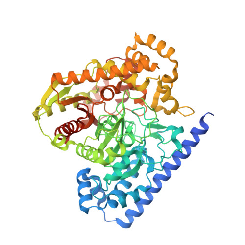
X-ray crystallographic analysis of alpha-ketoheterocycle inhibitors bound to a humanized variant of fatty acid amide hydrolase.

Mileni, M.Garfunkle, J.Ezzili, C.Kimball, F.S.Cravatt, B.F.Stevens, R.C.Boger, D.L.

(2010) J Med Chem 53: 230-240

  • DOI: https://doi.org/10.1021/jm9012196
  • Primary Citation Related Structures: 
    3K7F, 3K83, 3K84

  • PubMed Abstract: 

    Three cocrystal X-ray structures of the alpha-ketoheterocycle inhibitors 3-5 bound to a humanized variant of fatty acid amide hydrolase (FAAH) are disclosed and comparatively discussed alongside those of 1 (OL-135) and its isomer 2. These five X-ray structures systematically probe each of the three active site regions key to substrate or inhibitor binding: (1) the conformationally mobile acyl chain-binding pocket and membrane access channel responsible for fatty acid amide substrate and inhibitor acyl chain binding, (2) the atypical active site catalytic residues and surrounding oxyanion hole that covalently binds the core of the alpha-ketoheterocycle inhibitors captured as deprotonated hemiketals mimicking the tetrahedral intermediate of the enzyme-catalyzed reaction, and (3) the cytosolic port and its uniquely important imbedded ordered water molecules and a newly identified anion binding site. The detailed analysis of their key active site interactions and their implications on the interpretation of the available structure-activity relationships are discussed providing important insights for future design.


  • Organizational Affiliation
    • Department of Chemistry, The Skaggs Institute for Chemical Biology,The Scripps Research Institute, 10550 North Torrey Pines Road, La Jolla, California 92037, USA.

Macromolecule Content 

  • Total Structure Weight: 127.04 kDa 
  • Atom Count: 9,589 
  • Modeled Residue Count: 1,088 
  • Deposited Residue Count: 1,146 
  • Unique protein chains: 1

Macromolecules

Find similar proteins by:|  3D Structure
Entity ID: 1
MoleculeChains  Sequence LengthOrganismDetailsImage
Fatty-acid amide hydrolase 1
A, B
573Rattus norvegicusMutation(s): 6 
Gene Names: Faahfaah-1Faah1
EC: 3.5.1 (PDB Primary Data), 3.1.1 (UniProt), 3.5.1.99 (UniProt)
UniProt
Find proteins for P97612 (Rattus norvegicus)
Explore P97612 
Go to UniProtKB:  P97612
Entity Groups
Sequence Clusters30% Identity50% Identity70% Identity90% Identity95% Identity100% Identity
UniProt GroupP97612
Sequence Annotations
Expand
Reference Sequence

Small Molecules

Binding Affinity Annotations 
IDSourceBinding Affinity
F2C BindingDB:  3K7F Ki: min: 10.3, max: 20 (nM) from 3 assay(s)
IC50: 100 (nM) from 1 assay(s)

Experimental Data & Validation

Experimental Data

  • Method: X-RAY DIFFRACTION
  • Resolution: 1.95 Å
  • R-Value Free:  0.188 (Depositor), 0.186 (DCC) 
  • R-Value Work:  0.154 (Depositor), 0.153 (DCC) 
  • R-Value Observed: 0.156 (Depositor) 
Space Group: P 32 2 1
Unit Cell:
Length ( Å )Angle ( ˚ )
a = 103.87α = 90
b = 103.87β = 90
c = 255.41γ = 120
Software Package:
Software NamePurpose
XDSdata scaling
PHASERphasing
PHENIXrefinement
XDSdata reduction
XSCALEdata scaling

Structure Validation

View Full Validation Report


Ligand Structure Quality Assessment 


Entry History 

Deposition Data

Revision History  (Full details and data files)

  • Version 1.0: 2009-12-01
    Type: Initial release
  • Version 1.1: 2011-07-13
    Changes: Version format compliance
  • Version 1.2: 2021-10-13
    Changes: Database references, Derived calculations
  • Version 1.3: 2023-09-06
    Changes: Data collection, Refinement description
  • Version 1.4: 2024-10-30
    Changes: Structure summary