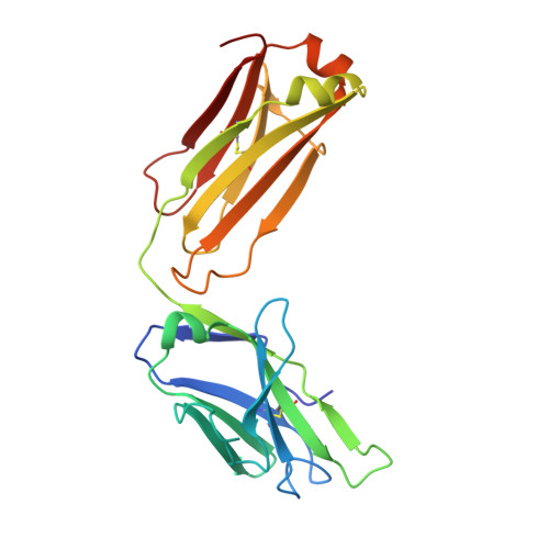
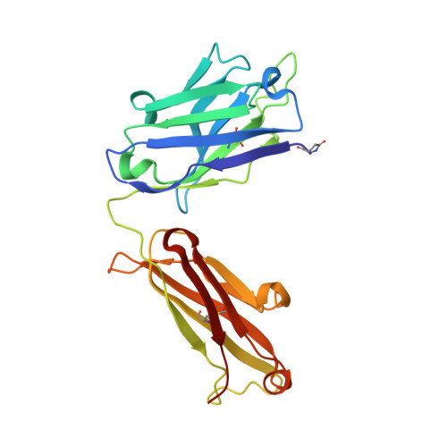
3KDM

Crystal Structure of Human Anti-steroid Fab 5F2 in Complex with Testosterone


Experimental Data Snapshot

  • Method: X-RAY DIFFRACTION
  • Resolution: 1.50 Å
  • R-Value Free: 0.208 
  • R-Value Work: 0.180 
  • R-Value Observed: 0.181 

Starting Model: experimental
View more details

wwPDB Validation   3D Report Full Report


Ligand Structure Quality Assessment 


This is version 2.1 of the entry. See complete history


Literature

The testosterone binding mechanism of an antibody derived from a naive human scFv library

Niemi, M.H.Takkinen, K.Amundsen, L.K.Soderlund, H.Rouvinen, J.Hoyhtya, M.

(2010) J Mol Recognit 24: 209-219

  • DOI: https://doi.org/10.1002/jmr.1039
  • Primary Citation of Related Structures:  
    3KDM

  • PubMed Abstract: 

    A testosterone binding scFv antibody was isolated from a naïve human library with a modest size of 10(8) clones. The crystal structure of the Fab fragment form of the 5F2 antibody clone complexed with testosterone determined at 1.5 Å resolution shows that the hapten is bound deeply in the antibody binding pocket. In addition to the interactions with framework residues only CDR-L3 and CDR-H3 loops interact with testosterone and the heavy chain forms the majority of the contacts with the hapten. The testosterone binding site of the 5F2 antibody with a high abundance of aromatic amino acid residues shows similarity with an in vitro affinity matured antibody having around 300 times higher affinity. The moderate affinity of the 5F2 antibody originates from the different orientation of the hapten and few light chain contacts. This is the first three-dimensional structure of a human steroid hormone binding antibody that has been isolated from a naïve human repertoire.


  • Organizational Affiliation

    Department of Chemistry, University of Eastern Finland, P.O. Box 111, FIN-80101 Joensuu, Finland.


Macromolecules
Find similar proteins by:  (by identity cutoff)  |  3D Structure
Entity ID: 1
MoleculeChains Sequence LengthOrganismDetailsImage
Immunoglobulin light chainA [auth L],
C [auth A]
218Homo sapiensMutation(s): 0 
Entity Groups  
Sequence Clusters30% Identity50% Identity70% Identity90% Identity95% Identity100% Identity
Sequence Annotations
Expand
  • Reference Sequence
Find similar proteins by:  (by identity cutoff)  |  3D Structure
Entity ID: 2
MoleculeChains Sequence LengthOrganismDetailsImage
Immunoglobulin heavy chainB [auth H],
D [auth B]
225Homo sapiensMutation(s): 0 
Entity Groups  
Sequence Clusters30% Identity50% Identity70% Identity90% Identity95% Identity100% Identity
Sequence Annotations
Expand
  • Reference Sequence
Small Molecules
Modified Residues  1 Unique
IDChains TypeFormula2D DiagramParent
PCA
Query on PCA
B [auth H],
D [auth B]
L-PEPTIDE LINKINGC5 H7 N O3GLN
Binding Affinity Annotations 
IDSourceBinding Affinity
TES PDBBind:  3KDM Kd: 100 (nM) from 1 assay(s)
Experimental Data & Validation

Experimental Data

  • Method: X-RAY DIFFRACTION
  • Resolution: 1.50 Å
  • R-Value Free: 0.208 
  • R-Value Work: 0.180 
  • R-Value Observed: 0.181 
  • Space Group: P 1
Unit Cell:
Length ( Å )Angle ( ˚ )
a = 66.75α = 81.32
b = 67.08β = 69.3
c = 67.06γ = 69.27
Software Package:
Software NamePurpose
PHENIXrefinement
PDB_EXTRACTdata extraction
ADSCdata collection
XDSdata reduction
XSCALEdata scaling
MOLREPphasing

Structure Validation

View Full Validation Report


Ligand Structure Quality Assessment 


Entry History 

Deposition Data

Revision History  (Full details and data files)

  • Version 1.0: 2010-05-26
    Type: Initial release
  • Version 1.1: 2011-07-13
    Changes: Version format compliance
  • Version 1.2: 2011-12-07
    Changes: Database references
  • Version 2.0: 2019-12-25
    Changes: Derived calculations, Polymer sequence
  • Version 2.1: 2023-11-01
    Changes: Data collection, Database references, Derived calculations, Refinement description