3KGD | pdb_00003kgd

Crystal structure of E. coli RNA 3' cyclase


Experimental Data Snapshot

  • Method: X-RAY DIFFRACTION
  • Resolution: 1.68 Å
  • R-Value Free: 
    0.207 (Depositor), 0.204 (DCC) 
  • R-Value Work: 
    0.168 (Depositor), 0.166 (DCC) 
  • R-Value Observed: 
    0.170 (Depositor) 

Starting Model: experimental
View more details

wwPDB Validation 3D Report Full Report

Validation slider image for 3KGD

Ligand Structure Quality Assessment 


This is version 1.5 of the entry. See complete history

Literature

Structure of the RNA 3'-phosphate cyclase-adenylate intermediate illuminates nucleotide specificity and covalent nucleotidyl transfer.

Tanaka, N.Smith, P.Shuman, S.

(2010) Structure 18: 449-457

  • DOI: https://doi.org/10.1016/j.str.2010.01.016
  • Primary Citation Related Structures: 
    3KGD

  • PubMed Abstract: 

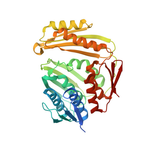
    RNA 3'-phosphate cyclase (RtcA) synthesizes RNA 2',3' cyclic phosphate ends via three steps: reaction with ATP to form a covalent RtcA-AMP intermediate; transfer of adenylate to an RNA 3'-phosphate to form RNA(3')pp(5')A; and attack of the vicinal O2' on the 3'-phosphorus to form a 2',3' cyclic phosphate. Here we report the 1.7 A crystal structure of the RtcA-AMP intermediate, which reveals the mechanism of nucleotidyl transfer. Adenylate is linked via a phosphoamide bond to the His309 Nepsilon atom. A network of hydrogen bonds to the ribose O2' and O3' accounts for the stringent ribonucleotide preference. Adenine is sandwiched in a hydrophobic pocket between Tyr284 and Pro131 and the preference for adenine is enforced by Phe135, which packs against the purine C2 edge. Two sulfates bound near the adenylate plausibly mimic the 3'-terminal and penultimate phosphates of RNA. The structure illuminates how the four alpha2/beta4 domains contribute to substrate binding and catalysis.


  • Organizational Affiliation
    • Molecular Biology Program, Sloan-Kettering Institute, New York, NY 10065, USA.

Macromolecule Content 

  • Total Structure Weight: 156.57 kDa 
  • Atom Count: 12,204 
  • Modeled Residue Count: 1,356 
  • Deposited Residue Count: 1,432 
  • Unique protein chains: 1

Macromolecules

Find similar proteins by:|  3D Structure
Entity ID: 1
MoleculeChains  Sequence LengthOrganismDetailsImage
RNA 3'-terminal phosphate cyclase
A, B, C, D
358Escherichia coli str. K-12 substr. MG1655Mutation(s): 0 
Gene Names: b4475JW5688rtcAyhgJyhgKYP_026219
EC: 6.5.1.4
UniProt
Find proteins for P46849 (Escherichia coli (strain K12))
Explore P46849 
Go to UniProtKB:  P46849
Entity Groups
Sequence Clusters30% Identity50% Identity70% Identity90% Identity95% Identity100% Identity
UniProt GroupP46849
Sequence Annotations
Expand
Reference Sequence

Small Molecules

Ligands 4 Unique
IDChains Name / Formula / InChI Key2D Diagram3D Interactions
AMP

Query on AMP


Download:Ideal Coordinates CCD File
E [auth A],
L [auth B],
R [auth C],
Z [auth D]
ADENOSINE MONOPHOSPHATE
C10 H14 N5 O7 P
UDMBCSSLTHHNCD-KQYNXXCUSA-N
SO4

Query on SO4


Download:Ideal Coordinates CCD File
EA [auth D]
I [auth A]
J [auth A]
K [auth A]
P [auth B]
EA [auth D],
I [auth A],
J [auth A],
K [auth A],
P [auth B],
Q [auth B],
X [auth C],
Y [auth C]
SULFATE ION
O4 S
QAOWNCQODCNURD-UHFFFAOYSA-L
GOL

Query on GOL


Download:Ideal Coordinates CCD File
AA [auth D]
BA [auth D]
CA [auth D]
F [auth A]
G [auth A]
AA [auth D],
BA [auth D],
CA [auth D],
F [auth A],
G [auth A],
M [auth B],
N [auth B],
S [auth C],
T [auth C],
U [auth C]
GLYCEROL
C3 H8 O3
PEDCQBHIVMGVHV-UHFFFAOYSA-N
NA

Query on NA


Download:Ideal Coordinates CCD File
DA [auth D],
H [auth A],
O [auth B],
V [auth C],
W [auth C]
SODIUM ION
Na
FKNQFGJONOIPTF-UHFFFAOYSA-N

Experimental Data & Validation

Experimental Data

  • Method: X-RAY DIFFRACTION
  • Resolution: 1.68 Å
  • R-Value Free:  0.207 (Depositor), 0.204 (DCC) 
  • R-Value Work:  0.168 (Depositor), 0.166 (DCC) 
  • R-Value Observed: 0.170 (Depositor) 
Space Group: P 1 21 1
Unit Cell:
Length ( Å )Angle ( ˚ )
a = 83.728α = 90
b = 81.932β = 103.44
c = 105.176γ = 90
Software Package:
Software NamePurpose
DENZOdata reduction
SCALEPACKdata scaling
PHASERphasing
PHENIXrefinement
PDB_EXTRACTdata extraction
HKL-2000data collection

Structure Validation

View Full Validation Report


Ligand Structure Quality Assessment 


Entry History 

Deposition Data

Revision History  (Full details and data files)

  • Version 1.0: 2010-04-21
    Type: Initial release
  • Version 1.1: 2011-07-13
    Changes: Source and taxonomy, Version format compliance
  • Version 1.2: 2012-03-21
    Changes: Database references
  • Version 1.3: 2017-11-01
    Changes: Refinement description
  • Version 1.4: 2023-09-06
    Changes: Data collection, Database references, Derived calculations, Refinement description
  • Version 1.5: 2024-11-06
    Changes: Structure summary