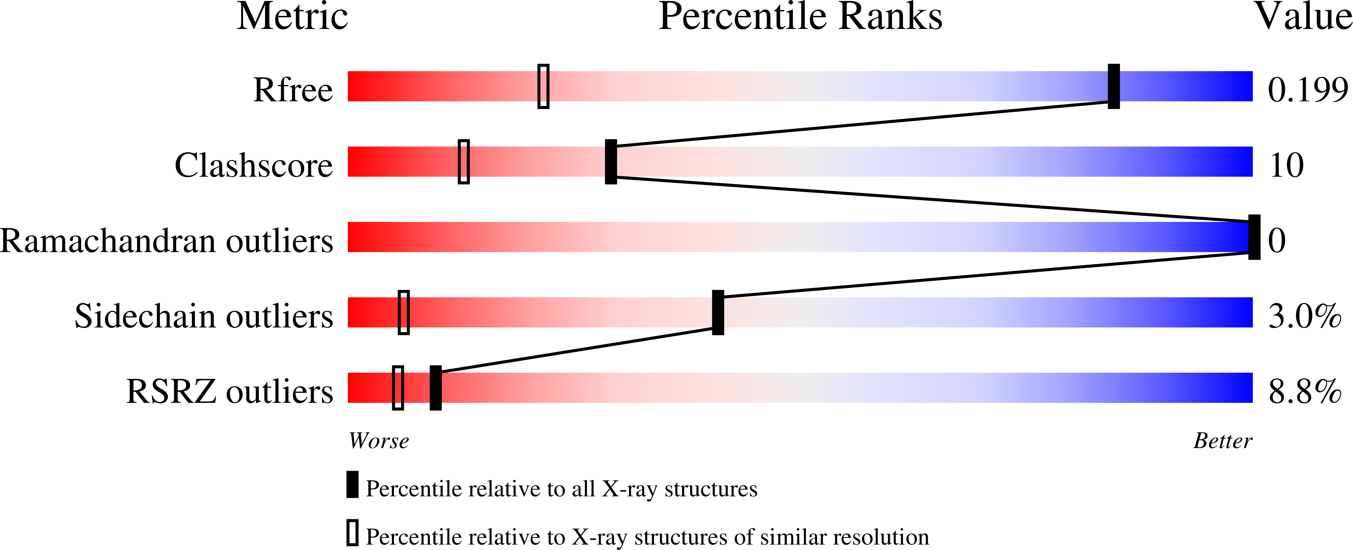
X-ray structure of the metcyano form of dehaloperoxidase from Amphitrite ornata: evidence for photoreductive dissociation of the iron-cyanide bond.
de Serrano, V.S., Davis, M.F., Gaff, J.F., Zhang, Q., Chen, Z., D'Antonio, E.L., Bowden, E.F., Rose, R., Franzen, S.(2010) Acta Crystallogr D Biol Crystallogr 66: 770-782
- PubMed: 20606257
- DOI: https://doi.org/10.1107/S0907444910014605
- Primary Citation of Related Structures:
3KUN, 3KUO - PubMed Abstract:
X-ray crystal structures of the metcyano form of dehaloperoxidase-hemoglobin (DHP A) from Amphitrite ornata (DHPCN) and the C73S mutant of DHP A (C73SCN) were determined using synchrotron radiation in order to further investigate the geometry of diatomic ligands coordinated to the heme iron. The DHPCN structure was also determined using a rotating-anode source. The structures show evidence of photoreduction of the iron accompanied by dissociation of bound cyanide ion (CN(-)) that depend on the intensity of the X-ray radiation and the exposure time. The electron density is consistent with diatomic molecules located in two sites in the distal pocket of DHPCN. However, the identities of the diatomic ligands at these two sites are not uniquely determined by the electron-density map. Consequently, density functional theory calculations were conducted in order to determine whether the bond lengths, angles and dissociation energies are consistent with bound CN(-) or O(2) in the iron-bound site. In addition, molecular-dynamics simulations were carried out in order to determine whether the dynamics are consistent with trapped CN(-) or O(2) in the second site of the distal pocket. Based on these calculations and comparison with a previously determined X-ray crystal structure of the C73S-O(2) form of DHP [de Serrano et al. (2007), Acta Cryst. D63, 1094-1101], it is concluded that CN(-) is gradually replaced by O(2) as crystalline DHP is photoreduced at 100 K. The ease of photoreduction of DHP A is consistent with the reduction potential, but suggests an alternative activation mechanism for DHP A compared with other peroxidases, which typically have reduction potentials that are 0.5 V more negative. The lability of CN(-) at 100 K suggests that the distal pocket of DHP A has greater flexibility than most other hemoglobins.
Organizational Affiliation:
Department of Chemistry, North Carolina State University, Raleigh, NC 27695, USA.


