


The Crystal Structure of beta-hexosaminidase B in complex with Pyrimethamine
Bateman, K.S., Cherney, M.M., Withers, S.G., Mahuran, D.J., Tropak, M., James, M.N.G.To be published.
Experimental Data Snapshot
Starting Model: experimental
View more details
Entity ID: 1 | |||||
|---|---|---|---|---|---|
| Molecule | Chains | Sequence Length | Organism | Details | Image |
| Beta-hexosaminidase subunit beta | 556 | Homo sapiens | Mutation(s): 0 EC: 3.2.1.52 | 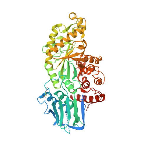 | |
UniProt & NIH Common Fund Data Resources | |||||
PHAROS: P07686 GTEx: ENSG00000049860 | |||||
Entity Groups | |||||
| Sequence Clusters | 30% Identity50% Identity70% Identity90% Identity95% Identity100% Identity | ||||
| UniProt Group | P07686 | ||||
Glycosylation | |||||
| Glycosylation Sites: 2 | Go to GlyGen: P07686-1 | ||||
Sequence AnnotationsExpand | |||||
Reference Sequence | |||||
Entity ID: 2 | |||||
|---|---|---|---|---|---|
| Molecule | Chains | Length | 2D Diagram | Glycosylation | D Interactions |
| beta-D-mannopyranose-(1-4)-2-acetamido-2-deoxy-beta-D-glucopyranose-(1-4)-2-acetamido-2-deoxy-beta-D-glucopyranose | C | 3 |  | N-Glycosylation | |
Glycosylation Resources | |||||
GlyTouCan: G15407YE GlyCosmos: G15407YE GlyGen: G15407YE | |||||
| Ligands 4 Unique | |||||
|---|---|---|---|---|---|
| ID | Chains | Name / Formula / InChI Key | 2D Diagram | 3D Interactions | |
| CP6 Download:Ideal Coordinates CCD File | G [auth A], L [auth B] | 5-(4-CHLORO-PHENYL)-6-ETHYL-PYRIMIDINE-2,4-DIAMINE C12 H13 Cl N4 WKSAUQYGYAYLPV-UHFFFAOYSA-N |  | ||
| NAG Download:Ideal Coordinates CCD File | F [auth A], H [auth B], I [auth B] | 2-acetamido-2-deoxy-beta-D-glucopyranose C8 H15 N O6 OVRNDRQMDRJTHS-FMDGEEDCSA-N |  | ||
| NDG Download:Ideal Coordinates CCD File | E [auth A], J [auth B] | 2-acetamido-2-deoxy-alpha-D-glucopyranose C8 H15 N O6 OVRNDRQMDRJTHS-PVFLNQBWSA-N |  | ||
| SO4 Download:Ideal Coordinates CCD File | K [auth B] | SULFATE ION O4 S QAOWNCQODCNURD-UHFFFAOYSA-L |  | ||
| Length ( Å ) | Angle ( ˚ ) |
|---|---|
| a = 113.898 | α = 90 |
| b = 113.898 | β = 90 |
| c = 397.427 | γ = 120 |
| Software Name | Purpose |
|---|---|
| Blu-Ice | data collection |
| CCP4 | model building |
| REFMAC | refinement |
| HKL-2000 | data reduction |
| HKL-2000 | data scaling |
| CCP4 | phasing |


