3LPP | pdb_00003lpp

Crystal complex of N-terminal sucrase-isomaltase with kotalanol


Experimental Data Snapshot

  • Method: X-RAY DIFFRACTION
  • Resolution: 2.15 Å
  • R-Value Free: 
    0.223 (Depositor), 0.274 (DCC) 
  • R-Value Work: 
    0.177 (Depositor), 0.240 (DCC) 
  • R-Value Observed: 
    0.179 (Depositor) 

wwPDB Validation   3D Report Full Report


Ligand Structure Quality Assessment 

Created with Raphaël 2.3.0Worse 01 BetterLigand structure goodness of fit to experimental dataBest fitted KTLClick on this verticalbar to view detailsBest fitted NAGClick on this verticalbar to view detailsBest fitted BMAClick on this verticalbar to view details

This is version 2.1 of the entry. See complete history


Literature

Structural basis for substrate selectivity in human maltase-glucoamylase and sucrase-isomaltase N-terminal domains.

Sim, L.Willemsma, C.Mohan, S.Naim, H.Y.Pinto, B.M.Rose, D.R.

(2010) J Biological Chem 285: 17763-17770

  • DOI: https://doi.org/10.1074/jbc.M109.078980
  • Primary Citation of Related Structures:  
    3LPO, 3LPP

  • PubMed Abstract: 

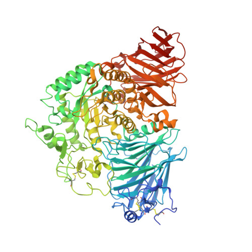
    Human maltase-glucoamylase (MGAM) and sucrase-isomaltase (SI) are small intestinal enzymes that work concurrently to hydrolyze the mixture of linear alpha-1,4- and branched alpha-1,6-oligosaccharide substrates that typically make up terminal starch digestion products. MGAM and SI are each composed of duplicated catalytic domains, N- and C-terminal, which display overlapping substrate specificities. The N-terminal catalytic domain of human MGAM (ntMGAM) has a preference for short linear alpha-1,4-oligosaccharides, whereas N-terminal SI (ntSI) has a broader specificity for both alpha-1,4- and alpha-1,6-oligosaccharides. Here we present the crystal structure of the human ntSI, in apo form to 3.2 A and in complex with the inhibitor kotalanol to 2.15 A resolution. Structural comparison with the previously solved structure of ntMGAM reveals key active site differences in ntSI, including a narrow hydrophobic +1 subsite, which may account for its additional substrate specificity for alpha-1,6 substrates.


  • Organizational Affiliation

    Department of Medical Biophysics, University of Toronto, Ontario Cancer Institute, Toronto, Ontario M56 2M9, Canada.


Macromolecules
Find similar proteins by:  (by identity cutoff)  |  3D Structure
Entity ID: 1
MoleculeChains Sequence LengthOrganismDetailsImage
Sucrase-isomaltase
A, B, C, D
898Homo sapiensMutation(s): 0 
Gene Names: SI
EC: 3.2.1.10
UniProt & NIH Common Fund Data Resources
Find proteins for P14410 (Homo sapiens)
Explore P14410 
Go to UniProtKB:  P14410
PHAROS:  P14410
GTEx:  ENSG00000090402 
Entity Groups  
Sequence Clusters30% Identity50% Identity70% Identity90% Identity95% Identity100% Identity
UniProt GroupP14410
Glycosylation
Glycosylation Sites: 4Go to GlyGen: P14410-1
Sequence Annotations
Expand
  • Reference Sequence
Oligosaccharides

Help

Entity ID: 2
MoleculeChains Length2D Diagram Glycosylation3D Interactions
alpha-D-mannopyranose-(1-3)-beta-D-mannopyranose-(1-4)-2-acetamido-2-deoxy-beta-D-glucopyranose-(1-4)-2-acetamido-2-deoxy-beta-D-glucopyranose
E, H
4N-Glycosylation
Glycosylation Resources
GlyTouCan:  G81315DD
GlyCosmos:  G81315DD
GlyGen:  G81315DD
Entity ID: 3
MoleculeChains Length2D Diagram Glycosylation3D Interactions
2-acetamido-2-deoxy-beta-D-glucopyranose-(1-4)-2-acetamido-2-deoxy-beta-D-glucopyranose
F, G, I
2N-Glycosylation
Glycosylation Resources
GlyTouCan:  G42666HT
GlyCosmos:  G42666HT
GlyGen:  G42666HT
Small Molecules
Ligands 6 Unique
IDChains Name / Formula / InChI Key2D Diagram3D Interactions
KTL
Query on KTL

Download Ideal Coordinates CCD File 
Q [auth B],
X [auth D]
(1S,2R,3R,4S)-1-{(1S)-2-[(2R,3S,4S)-3,4-dihydroxy-2-(hydroxymethyl)tetrahydrothiophenium-1-yl]-1-hydroxyethyl}-2,3,4,5-tetrahydroxypentyl sulfate
C12 H24 O12 S2
OMKXVFDVAGCPBS-QTIKZJLFSA-N
NAG
Query on NAG

Download Ideal Coordinates CCD File 
J [auth A]
K [auth A]
O [auth B]
S [auth C]
T [auth C]
2-acetamido-2-deoxy-beta-D-glucopyranose
C8 H15 N O6
OVRNDRQMDRJTHS-FMDGEEDCSA-N
BMA
Query on BMA

Download Ideal Coordinates CCD File 
P [auth B]beta-D-mannopyranose
C6 H12 O6
WQZGKKKJIJFFOK-RWOPYEJCSA-N
TRS
Query on TRS

Download Ideal Coordinates CCD File 
L [auth A],
V [auth C]
2-AMINO-2-HYDROXYMETHYL-PROPANE-1,3-DIOL
C4 H12 N O3
LENZDBCJOHFCAS-UHFFFAOYSA-O
PEG
Query on PEG

Download Ideal Coordinates CCD File 
M [auth A],
R [auth B],
W [auth C],
Y [auth D]
DI(HYDROXYETHYL)ETHER
C4 H10 O3
MTHSVFCYNBDYFN-UHFFFAOYSA-N
CL
Query on CL

Download Ideal Coordinates CCD File 
N [auth A]CHLORIDE ION
Cl
VEXZGXHMUGYJMC-UHFFFAOYSA-M
Experimental Data & Validation

Experimental Data

  • Method: X-RAY DIFFRACTION
  • Resolution: 2.15 Å
  • R-Value Free:  0.223 (Depositor), 0.274 (DCC) 
  • R-Value Work:  0.177 (Depositor), 0.240 (DCC) 
  • R-Value Observed: 0.179 (Depositor) 
Space Group: P 21 21 21
Unit Cell:
Length ( Å )Angle ( ˚ )
a = 69.403α = 90
b = 165.763β = 90
c = 341.4γ = 90
Software Package:
Software NamePurpose
DENZOdata reduction
SCALEPACKdata scaling
PHASERphasing
REFMACrefinement
PDB_EXTRACTdata extraction

Structure Validation

View Full Validation Report


Ligand Structure Quality Assessment 

Created with Raphaël 2.3.0Worse 01 BetterLigand structure goodness of fit to experimental dataBest fitted KTLClick on this verticalbar to view detailsBest fitted NAGClick on this verticalbar to view detailsBest fitted BMAClick on this verticalbar to view details

Entry History 

Deposition Data

Revision History  (Full details and data files)

  • Version 1.0: 2010-03-31
    Type: Initial release
  • Version 1.1: 2011-07-13
    Changes: Version format compliance
  • Version 1.2: 2017-11-01
    Changes: Advisory, Refinement description
  • Version 2.0: 2020-07-29
    Type: Remediation
    Reason: Carbohydrate remediation
    Changes: Advisory, Atomic model, Data collection, Database references, Derived calculations, Structure summary
  • Version 2.1: 2024-10-30
    Changes: Data collection, Database references, Structure summary