3LT5

X-ray Crystallographic structure of a Pseudomonas Aeruginosa Azoreductase in complex with balsalazide


Experimental Data Snapshot

  • Method: X-RAY DIFFRACTION
  • Resolution: 2.30 Å
  • R-Value Free: 
    0.222 (Depositor), 0.220 (DCC) 
  • R-Value Work: 
    0.154 (Depositor), 0.150 (DCC) 
  • R-Value Observed: 
    0.157 (Depositor) 

Starting Model: experimental
View more details

wwPDB Validation   3D Report Full Report


Ligand Structure Quality Assessment 

Created with Raphaël 2.3.0Worse 01 BetterLigand structure goodness of fit to experimental dataBest fitted FMNClick on this verticalbar to view detailsBest fitted BLQClick on this verticalbar to view details

This is version 1.3 of the entry. See complete history


Literature

A Novel Mechanism for Azoreduction

Ryan, A.Laurieri, N.Westwood, I.Wang, C.-J.Lowe, E.Sim, E.

(2010) J Mol Biol 400: 24-37

  • DOI: https://doi.org/10.1016/j.jmb.2010.04.023
  • Primary Citation of Related Structures:  
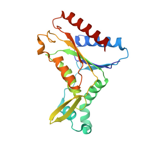
    3LT5

  • PubMed Abstract: 

    Azoreductases are important due to their ability to activate anti-inflammatory azo pro-drugs and to detoxify azo dyes. Three genes encoding azoreductases have been identified in Pseudomonas aeruginosa. We describe here a comparison of the three enzymes. The pure recombinant proteins each have a distinct substrate specificity profile against a range of azo substrates. Using the structure of P. aeruginosa azoreductase (paAzoR) 1 and the homology models of paAzoR2 and paAzoR3, we have identified residues important for substrate specificity. We have defined a novel flavin mononucleotide binding cradle, which is a recurrent motif in many flavodoxin-like proteins. A novel structure of paAzoR1 with the azo pro-drug balsalazide bound within the active site was determined by X-ray crystallography and demonstrates that the substrate is present in a hydrazone tautomer conformation. We propose that the structure with balsalazide bound represents an enzyme intermediate and, together with the flavin mononucleotide binding cradle, we propose a novel catalytic mechanism.


  • Organizational Affiliation

    Department of Pharmacology, University of Oxford, Mansfield Road, Oxford OX1 3QT, UK.


Macromolecules
Find similar proteins by:  (by identity cutoff)  |  3D Structure
Entity ID: 1
MoleculeChains Sequence LengthOrganismDetailsImage
FMN-dependent NADH-azoreductase 1
A, B
212Pseudomonas aeruginosa PAO1Mutation(s): 0 
Gene Names: PAO785
EC: 1.7 (PDB Primary Data), 1.6.5 (UniProt), 1.7.1.17 (UniProt)
UniProt
Find proteins for Q9I5F3 (Pseudomonas aeruginosa (strain ATCC 15692 / DSM 22644 / CIP 104116 / JCM 14847 / LMG 12228 / 1C / PRS 101 / PAO1))
Explore Q9I5F3 
Go to UniProtKB:  Q9I5F3
Entity Groups  
Sequence Clusters30% Identity50% Identity70% Identity90% Identity95% Identity100% Identity
UniProt GroupQ9I5F3
Sequence Annotations
Expand
  • Reference Sequence
Small Molecules
Ligands 3 Unique
IDChains Name / Formula / InChI Key2D Diagram3D Interactions
FMN
Query on FMN

Download Ideal Coordinates CCD File 
C [auth A],
G [auth B]
FLAVIN MONONUCLEOTIDE
C17 H21 N4 O9 P
FVTCRASFADXXNN-SCRDCRAPSA-N
BLQ
Query on BLQ

Download Ideal Coordinates CCD File 
E [auth A],
F [auth A]
(3E)-3-({4-[(2-carboxyethyl)carbamoyl]phenyl}hydrazono)-6-oxocyclohexa-1,4-diene-1-carboxylic acid
C17 H15 N3 O6
KONZVQJABTUMFX-UDWIEESQSA-N
GOL
Query on GOL

Download Ideal Coordinates CCD File 
D [auth A],
H [auth B],
I [auth B]
GLYCEROL
C3 H8 O3
PEDCQBHIVMGVHV-UHFFFAOYSA-N
Experimental Data & Validation

Experimental Data

  • Method: X-RAY DIFFRACTION
  • Resolution: 2.30 Å
  • R-Value Free:  0.222 (Depositor), 0.220 (DCC) 
  • R-Value Work:  0.154 (Depositor), 0.150 (DCC) 
  • R-Value Observed: 0.157 (Depositor) 
Space Group: P 31 2 1
Unit Cell:
Length ( Å )Angle ( ˚ )
a = 83.097α = 90
b = 83.097β = 90
c = 109.05γ = 120
Software Package:
Software NamePurpose
MOSFLMdata reduction
SCALAdata scaling
PHENIXrefinement
PDB_EXTRACTdata extraction

Structure Validation

View Full Validation Report


Ligand Structure Quality Assessment 

Created with Raphaël 2.3.0Worse 01 BetterLigand structure goodness of fit to experimental dataBest fitted FMNClick on this verticalbar to view detailsBest fitted BLQClick on this verticalbar to view details

Entry History 

Deposition Data

Revision History  (Full details and data files)

  • Version 1.0: 2010-05-12
    Type: Initial release
  • Version 1.1: 2011-07-13
    Changes: Version format compliance
  • Version 1.2: 2019-12-25
    Changes: Advisory, Data collection
  • Version 1.3: 2023-11-01
    Changes: Advisory, Data collection, Database references, Derived calculations, Refinement description