3MH7 | pdb_00003mh7

HtrA proteases are activated by a conserved mechanism that can be triggered by distinct molecular cues


Experimental Data Snapshot

  • Method: X-RAY DIFFRACTION
  • Resolution: 2.96 Å
  • R-Value Free: 
    0.249 (Depositor), 0.239 (DCC) 
  • R-Value Work: 
    0.199 (Depositor), 0.194 (DCC) 
  • R-Value Observed: 
    0.201 (Depositor) 

wwPDB Validation 3D Report Full Report

Validation slider image for 3MH7

This is version 1.3 of the entry. See complete history

Literature

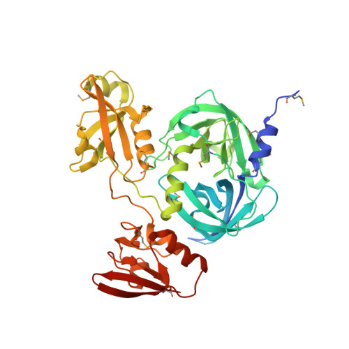
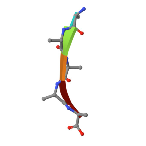
HtrA proteases have a conserved activation mechanism that can be triggered by distinct molecular cues

Krojer, T.Sawa, J.Huber, R.Clausen, T.

(2010) Nat Struct Mol Biol 17: 844-852

  • DOI: https://doi.org/10.1038/nsmb.1840
  • Primary Citation Related Structures: 
    3MH4, 3MH5, 3MH6, 3MH7

  • PubMed Abstract: 

    HtrA proteases are tightly regulated proteolytic assemblies that are essential for maintaining protein homeostasis in extracytosolic compartments. Though HtrA proteases have been characterized in detail, their precise molecular mechanism for switching between different functional states is still unknown. To address this, we carried out biochemical and structural studies of DegP from Escherichia coli. We show that effector-peptide binding to the PDZ domain of DegP induces oligomer conversion from resting hexameric DegP6 into proteolytically active 12-mers and 24-mers (DegP12/24). Moreover, our data demonstrate that a specific protease loop (L3) functions as a conserved molecular switch of HtrA proteases. L3 senses the activation signal-that is, the repositioned PDZ domain of substrate-engaged DegP12/24 or the binding of allosteric effectors to regulatory HtrA proteases such as DegS-and transmits this information to the active site. Implications for protein quality control and regulation of oligomeric enzymes are discussed.


  • Organizational Affiliation
    • Research Institute of Molecular Pathology, Vienna, Austria.

Macromolecule Content 

  • Total Structure Weight: 49.47 kDa 
  • Atom Count: 2,919 
  • Modeled Residue Count: 402 
  • Deposited Residue Count: 466 
  • Unique protein chains: 2

Macromolecules

Find similar proteins by:|  3D Structure
Entity ID: 1
MoleculeChains  Sequence LengthOrganismDetailsImage
Protease do456Escherichia coli K-12Mutation(s): 1 
Gene Names: DegP
EC: 3.4.21 (PDB Primary Data), 3.4.21.107 (UniProt)
UniProt
Find proteins for P0C0V0 (Escherichia coli (strain K12))
Explore P0C0V0 
Go to UniProtKB:  P0C0V0
Entity Groups
Sequence Clusters30% Identity50% Identity70% Identity90% Identity95% Identity100% Identity
UniProt GroupP0C0V0
Sequence Annotations
Expand
Reference Sequence
Find similar proteins by:  Sequence   |   3D Structure  
Entity ID: 2
MoleculeChains  Sequence LengthOrganismDetailsImage
5-mer peptide
B, C
5Escherichia coli K-12Mutation(s): 0 
Gene Names: DegP

Small Molecules

Modified Residues  1 Unique
IDChains TypeFormula2D DiagramParent
MSE
Query on MSE
A
L-PEPTIDE LINKINGC5 H11 N O2 SeMET
UNK
Query on UNK
B, C
L-PEPTIDE LINKINGC4 H9 N O2

--

Experimental Data & Validation

Experimental Data

  • Method: X-RAY DIFFRACTION
  • Resolution: 2.96 Å
  • R-Value Free:  0.249 (Depositor), 0.239 (DCC) 
  • R-Value Work:  0.199 (Depositor), 0.194 (DCC) 
  • R-Value Observed: 0.201 (Depositor) 
Space Group: F 4 3 2
Unit Cell:
Length ( Å )Angle ( ˚ )
a = 253.929α = 90
b = 253.929β = 90
c = 253.929γ = 90
Software Package:
Software NamePurpose
DNAdata collection
SHARPphasing
PHENIXrefinement
XDSdata reduction
XSCALEdata scaling

Structure Validation

View Full Validation Report


Entry History 

Deposition Data

Revision History  (Full details and data files)

  • Version 1.0: 2010-06-30
    Type: Initial release
  • Version 1.1: 2011-07-13
    Changes: Version format compliance
  • Version 1.2: 2014-03-05
    Changes: Database references
  • Version 1.3: 2024-10-30
    Changes: Data collection, Database references, Derived calculations, Structure summary