3N1C | pdb_00003n1c

Crystal structure of the phosphofructokinase-2 from Escherichia coli in complex with fructose-6-phosphate


Experimental Data Snapshot

  • Method: X-RAY DIFFRACTION
  • Resolution: 2.00 Å
  • R-Value Free: 
    0.221 (Depositor), 0.216 (DCC) 
  • R-Value Work: 
    0.180 (Depositor), 0.176 (DCC) 
  • R-Value Observed: 
    0.182 (Depositor) 

Starting Model: experimental
View more details

wwPDB Validation 3D Report Full Report

Validation slider image for 3N1C

Ligand Structure Quality Assessment 


This is version 1.3 of the entry. See complete history

Literature

The Crystal Complex of Phosphofructokinase-2 of Escherichia coli with Fructose-6-phosphate: KINETIC AND STRUCTURAL ANALYSIS OF THE ALLOSTERIC ATP INHIBITION.

Cabrera, R.Baez, M.Pereira, H.M.Caniuguir, A.Garratt, R.C.Babul, J.

(2011) J Biological Chem 286: 5774-5783

  • DOI: https://doi.org/10.1074/jbc.M110.163162
  • Primary Citation Related Structures: 
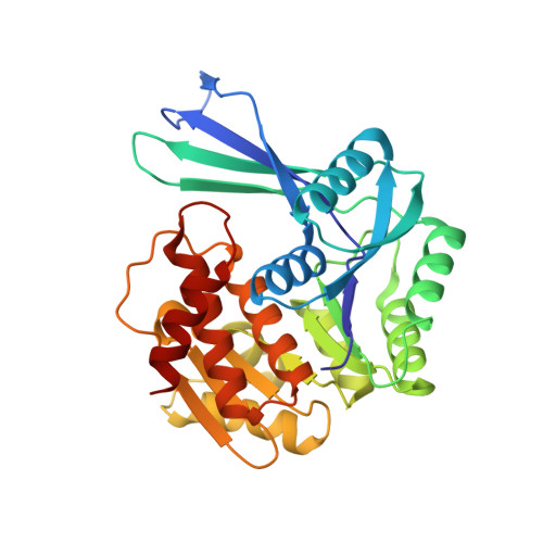
    3N1C

  • PubMed Abstract: 

    Substrate inhibition by ATP is a regulatory feature of the phosphofructokinases isoenzymes from Escherichia coli (Pfk-1 and Pfk-2). Under gluconeogenic conditions, the loss of this regulation in Pfk-2 causes substrate cycling of fructose-6-phosphate (fructose-6-P) and futile consumption of ATP delaying growth. In the present work, we have broached the mechanism of ATP-induced inhibition of Pfk-2 from both structural and kinetic perspectives. The crystal structure of Pfk-2 in complex with fructose-6-P is reported to a resolution of 2 Å. The comparison of this structure with the previously reported inhibited form of the enzyme suggests a negative interplay between fructose-6-P binding and allosteric binding of MgATP. Initial velocity experiments show a linear increase of the apparent K(0.5) for fructose-6-P and a decrease in the apparent k(cat) as a function of MgATP concentration. These effects occur simultaneously with the induction of a sigmoidal kinetic behavior (n(H) of approximately 2). Differences and resemblances in the patterns of fructose-6-P binding and the mechanism of inhibition are discussed for Pfk-1 and Pfk-2, as an example of evolutionary convergence, because these enzymes do not share a common ancestor.


  • Organizational Affiliation
    • Departamento de Biología, Facultad de Ciencias, Universidad de Chile, Santiago, Chile. ricabrer@uchile.cl

Macromolecule Content 

  • Total Structure Weight: 130.99 kDa 
  • Atom Count: 10,098 
  • Modeled Residue Count: 1,230 
  • Deposited Residue Count: 1,236 
  • Unique protein chains: 1

Macromolecules

Find similar proteins by:|  3D Structure
Entity ID: 1
MoleculeChains  Sequence LengthOrganismDetailsImage
6-phosphofructokinase isozyme 2
A, B, C, D
309Escherichia coli K-12Mutation(s): 0 
Gene Names: b1723JW5280PFK2pfkB
EC: 2.7.1.11
UniProt
Find proteins for P06999 (Escherichia coli (strain K12))
Explore P06999 
Go to UniProtKB:  P06999
Entity Groups
Sequence Clusters30% Identity50% Identity70% Identity90% Identity95% Identity100% Identity
UniProt GroupP06999
Sequence Annotations
Expand
Reference Sequence

Experimental Data & Validation

Experimental Data

  • Method: X-RAY DIFFRACTION
  • Resolution: 2.00 Å
  • R-Value Free:  0.221 (Depositor), 0.216 (DCC) 
  • R-Value Work:  0.180 (Depositor), 0.176 (DCC) 
  • R-Value Observed: 0.182 (Depositor) 
Space Group: C 2 2 21
Unit Cell:
Length ( Å )Angle ( ˚ )
a = 68.793α = 90
b = 153.605β = 90
c = 223.79γ = 90
Software Package:
Software NamePurpose
MOSFLMdata reduction
SCALAdata scaling
PHASERphasing
PHENIXrefinement
PDB_EXTRACTdata extraction

Structure Validation

View Full Validation Report


Ligand Structure Quality Assessment 


Entry History 

Deposition Data

Revision History  (Full details and data files)

  • Version 1.0: 2010-12-08
    Type: Initial release
  • Version 1.1: 2011-07-13
    Changes: Version format compliance
  • Version 1.2: 2020-07-29
    Type: Remediation
    Reason: Carbohydrate remediation
    Changes: Advisory, Data collection, Derived calculations, Structure summary
  • Version 1.3: 2023-09-06
    Changes: Data collection, Database references, Refinement description, Structure summary