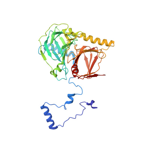
Crystal structures of salicylate 1,2-dioxygenase-substrates adducts: A step towards the comprehension of the structural basis for substrate selection in class III ring cleaving dioxygenases.
Ferraroni, M., Matera, I., Steimer, L., Burger, S., Scozzafava, A., Stolz, A., Briganti, F.(2012) J Struct Biol 177: 431-438
- PubMed: 22155290
- DOI: https://doi.org/10.1016/j.jsb.2011.11.026
- Primary Citation of Related Structures:
3NJZ, 3NKT, 3NL1 - PubMed Abstract:
The crystallographic structures of the adducts of salicylate 1,2-dioxygenase (SDO) with substrates salicylate, gentisate and 1-hydroxy-2-naphthoate, obtained under anaerobic conditions, have been solved and analyzed. This ring fission dioxygenase from the naphthalenesulfonate-degrading bacterium Pseudaminobacter salicylatoxidans BN12, is a homo-tetrameric class III ring-cleaving dioxygenase containing a catalytic Fe(II) ion coordinated by three histidine residues. SDO is markedly different from the known gentisate 1,2-dioxygenases or 1-hydroxy-2-naphthoate dioxygenases, belonging to the same class, because of its unique ability to oxidatively cleave salicylate, gentisate and 1-hydroxy-2-naphthoate. The crystal structures of the anaerobic complexes of the SDO reveal the mode of binding of the substrates into the active site and unveil the residues which are important for the correct positioning of the substrate molecules. Upon binding of the substrates the active site of SDO undergoes a series of conformational changes: in particular Arg127, His162, and Arg83 move to make hydrogen bond interactions with the carboxyl group of the substrate molecules. Unpredicted concerted displacements upon substrate binding are observed for the loops composed of residues 40-43, 75-85, and 192-198 where several aminoacidic residues, such as Leu42, Arg79, Arg83, and Asp194, contribute to the closing of the active site together with the amino-terminal tail (residues 2-15). Differences in substrate specificity are controlled by several residues located in the upper part of the substrate binding cavity like Met46, Ala85, Trp104, and Phe189, although we cannot exclude that the kinetic differences observed could also be generated by concerted conformational changes resulting from amino-acid mutations far from the active site.
- Dipartimento di Chimica Ugo Schiff, Università di Firenze, Via della Lastruccia 3, I-50019 Sesto Fiorentino (FI), Italy.
Organizational Affiliation:


