3PX2 | pdb_00003px2

Structure of TylM1 from Streptomyces fradiae H123N mutant in complex with SAH and dTDP-Quip3N


Experimental Data Snapshot

  • Method: X-RAY DIFFRACTION
  • Resolution: 1.65 Å
  • R-Value Free: 
    0.245 (Depositor), 0.240 (DCC) 
  • R-Value Work: 
    0.194 (Depositor), 0.190 (DCC) 
  • R-Value Observed: 
    0.197 (Depositor) 

Starting Model: experimental
View more details

wwPDB Validation 3D Report Full Report

Validation slider image for 3PX2

Ligand Structure Quality Assessment 


This is version 1.2 of the entry. See complete history

Literature

Molecular Architecture of TylM1 from Streptomyces fradiae: An N,N-Dimethyltransferase Involved in the Production of dTDP-d-mycaminose .

Carney, A.E.Holden, H.M.

(2011) Biochemistry 50: 780-787

  • DOI: https://doi.org/10.1021/bi101733y
  • Primary Citation Related Structures: 
    3PFG, 3PFH, 3PX2, 3PX3

  • PubMed Abstract: 

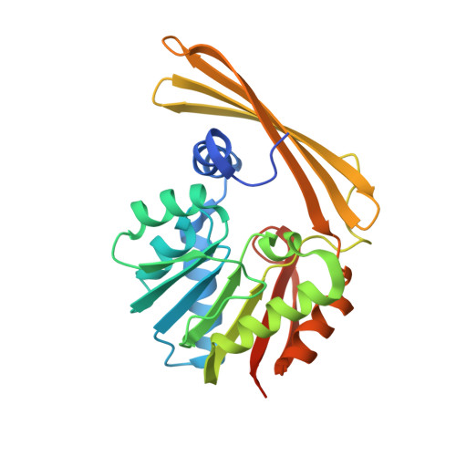
    d-Mycaminose is an unusual dideoxy sugar found attached to the antibiotic tylosin, a commonly used veterinarian therapeutic. It is synthesized by the Gram-positive bacterium Streptomyces fradiae as a dTDP-linked sugar. The last step in its biosynthesis involves the dimethylation of the hexose C-3' amino group by an S-adenosylmethionine (SAM) dependent enzyme referred to as TylM1. Here we report two high-resolution X-ray structures of TylM1, one in which the enzyme contains bound SAM and dTDP-phenol and the second in which the protein is complexed with S-adenosylhomocysteine (SAH) and dTDP-3-amino-3,6-dideoxyglucose, its natural substrate. Combined, these two structures, solved to 1.35 and 1.79 Å resolution, respectively, show the orientations of SAM and the dTDP-linked sugar substrate within the active site region. Specifically, the C-3' amino group of the hexose is in the correct position for an in-line attack at the reactive methyl group of SAM. Both Tyr 14 and Arg 241 serve to anchor the dTDP-linked sugar to the protein. To test the role of His 123 in catalysis, two site-directed mutant proteins were constructed, H123A and H123N. Both mutant proteins retained catalytic activity, albeit with reduced rates. Specifically, the k(cat)/K(m) was reduced to 1.8% and 0.37% for the H123A and H123N mutant proteins, respectively. High-resolution X-ray models showed that the observed perturbations in the kinetic constants were not due to major changes in their three-dimensional folds. Most likely the proton on the C-3' amino group is transferred to one of the water molecules lining the active site pocket as catalysis proceeds.


  • Organizational Affiliation
    • Department of Biochemistry, University of Wisconsin, Madison, Wisconsin 53706, United States.

Macromolecule Content 

  • Total Structure Weight: 58.74 kDa 
  • Atom Count: 4,232 
  • Modeled Residue Count: 480 
  • Deposited Residue Count: 524 
  • Unique protein chains: 1

Macromolecules

Find similar proteins by:|  3D Structure
Entity ID: 1
MoleculeChains  Sequence LengthOrganismDetailsImage
N-methyltransferaseA,
B [auth D]
262Streptomyces fradiaeMutation(s): 1 
Gene Names: tylM1tylMI(orf3*)
EC: 2.1.1.235
UniProt
Find proteins for P95748 (Streptomyces fradiae)
Explore P95748 
Go to UniProtKB:  P95748
Entity Groups
Sequence Clusters30% Identity50% Identity70% Identity90% Identity95% Identity100% Identity
UniProt GroupP95748
Sequence Annotations
Expand
Reference Sequence

Small Molecules

Ligands 3 Unique
IDChains Name / Formula / InChI Key2D Diagram3D Interactions
T3Q

Query on T3Q


Download:Ideal Coordinates CCD File
D [auth A],
F [auth D]
[(3R,4S,5S,6R)-4-amino-3,5-dihydroxy-6-methyloxan-2-yl][hydroxy-[[(2R,3S,5R)-3-hydroxy-5-(5-methyl-2,4-dioxopyrimidin-1-yl)oxolan-2-yl]methoxy]phosphoryl] hydrogen phosphate
C16 H27 N3 O14 P2
KVYJLJOGNUNRJK-HALQBZCBSA-N
SAH

Query on SAH


Download:Ideal Coordinates CCD File
C [auth A],
E [auth D]
S-ADENOSYL-L-HOMOCYSTEINE
C14 H20 N6 O5 S
ZJUKTBDSGOFHSH-WFMPWKQPSA-N
EDO

Query on EDO


Download:Ideal Coordinates CCD File
G [auth D],
H [auth D]
1,2-ETHANEDIOL
C2 H6 O2
LYCAIKOWRPUZTN-UHFFFAOYSA-N

Experimental Data & Validation

Experimental Data

  • Method: X-RAY DIFFRACTION
  • Resolution: 1.65 Å
  • R-Value Free:  0.245 (Depositor), 0.240 (DCC) 
  • R-Value Work:  0.194 (Depositor), 0.190 (DCC) 
  • R-Value Observed: 0.197 (Depositor) 
Space Group: P 1 21 1
Unit Cell:
Length ( Å )Angle ( ˚ )
a = 86.571α = 90
b = 40.703β = 117.93
c = 87.208γ = 90
Software Package:
Software NamePurpose
PROTEUM PLUSdata collection
PHASERphasing
REFMACrefinement
SAINTdata reduction
SADABSdata scaling

Structure Validation

View Full Validation Report


Ligand Structure Quality Assessment 


Entry History 

Deposition Data

Revision History  (Full details and data files)

  • Version 1.0: 2010-12-22
    Type: Initial release
  • Version 1.1: 2011-07-13
    Changes: Version format compliance
  • Version 1.2: 2023-09-13
    Changes: Data collection, Database references, Derived calculations, Refinement description