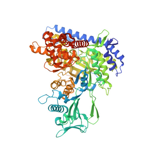
3S9I

Crystal structure of Mycobacterium tuberculosis malate synthase in complex with 2-4-dioxo-4-phenylbutanoic acid inhibitor


Experimental Data Snapshot

  • Method: X-RAY DIFFRACTION
  • Resolution: 1.90 Å
  • R-Value Free: 0.203 
  • R-Value Work: 0.168 
  • R-Value Observed: 0.170 

Starting Model: experimental
View more details

wwPDB Validation   3D Report Full Report


Ligand Structure Quality Assessment 


This is version 1.3 of the entry. See complete history


Literature

Structure-guided discovery of phenyl-diketo acids as potent inhibitors of M. tuberculosis malate synthase.

Krieger, I.V.Freundlich, J.S.Gawandi, V.B.Roberts, J.P.Gawandi, V.B.Sun, Q.Owen, J.L.Fraile, M.T.Huss, S.I.Lavandera, J.L.Ioerger, T.R.Sacchettini, J.C.

(2012) Chem Biol 19: 1556-1567

  • DOI: https://doi.org/10.1016/j.chembiol.2012.09.018
  • Primary Citation of Related Structures:  
    3S9I, 3S9Z, 3SAD, 3SAZ, 3SB0

  • PubMed Abstract: 

    The glyoxylate shunt plays an important role in fatty acid metabolism and has been shown to be critical to survival of several pathogens involved in chronic infections. For Mycobacterium tuberculosis (Mtb), a strain with a defective glyoxylate shunt was previously shown to be unable to establish infection in a mouse model. We report the development of phenyl-diketo acid (PDKA) inhibitors of malate synthase (GlcB), one of two glyoxylate shunt enzymes, using structure-based methods. PDKA inhibitors were active against Mtb grown on acetate, and overexpression of GlcB ameliorated this inhibition. Crystal structures of complexes of GlcB with PDKA inhibitors guided optimization of potency. A selected PDKA compound demonstrated efficacy in a mouse model of tuberculosis. The discovery of these PDKA derivatives provides chemical validation of GlcB as an attractive target for tuberculosis therapeutics.


  • Organizational Affiliation

    Department of Biochemistry and Biophysics, Texas A&M University, College Station, TX 77843, USA.


Macromolecules
Find similar proteins by:  (by identity cutoff)  |  3D Structure
Entity ID: 1
MoleculeChains Sequence LengthOrganismDetailsImage
Malate synthase G741Mycobacterium tuberculosisMutation(s): 1 
Gene Names: glcBMT1885MTCY1A11.06Rv1837c
EC: 2.3.3.9
UniProt
Find proteins for P9WK17 (Mycobacterium tuberculosis (strain ATCC 25618 / H37Rv))
Explore P9WK17 
Go to UniProtKB:  P9WK17
Entity Groups  
Sequence Clusters30% Identity50% Identity70% Identity90% Identity95% Identity100% Identity
UniProt GroupP9WK17
Sequence Annotations
Expand
  • Reference Sequence
Experimental Data & Validation

Experimental Data

  • Method: X-RAY DIFFRACTION
  • Resolution: 1.90 Å
  • R-Value Free: 0.203 
  • R-Value Work: 0.168 
  • R-Value Observed: 0.170 
  • Space Group: P 43 21 2
Unit Cell:
Length ( Å )Angle ( ˚ )
a = 79.331α = 90
b = 79.331β = 90
c = 225.945γ = 90
Software Package:
Software NamePurpose
HKL-2000data collection
BUSTERrefinement
HKL-2000data reduction
HKL-2000data scaling

Structure Validation

View Full Validation Report


Ligand Structure Quality Assessment 


Entry History 

Deposition Data

Revision History  (Full details and data files)

  • Version 1.0: 2012-11-07
    Type: Initial release
  • Version 1.1: 2013-02-20
    Changes: Database references
  • Version 1.2: 2017-11-08
    Changes: Refinement description
  • Version 1.3: 2023-09-13
    Changes: Data collection, Database references, Derived calculations, Refinement description