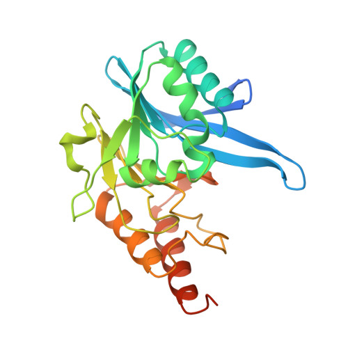
3SPU

apo NDM-1 Crystal Structure


Experimental Data Snapshot

  • Method: X-RAY DIFFRACTION
  • Resolution: 2.10 Å
  • R-Value Free: 0.253 
  • R-Value Work: 0.216 
  • R-Value Observed: 0.218 

Starting Model: experimental
View more details

wwPDB Validation   3D Report Full Report


This is version 2.1 of the entry. See complete history


Literature

Crystal structure of New Delhi metallo-beta-lactamase reveals molecular basis for antibiotic resistance

King, D.Strynadka, N.

(2011) Protein Sci 20: 1484-1491

  • DOI: https://doi.org/10.1002/pro.697
  • Primary Citation of Related Structures:  
    3SPU

  • PubMed Abstract: 

    β-Lactams are the most commonly prescribed class of antibiotics and have had an enormous impact on human health. Thus, it is disquieting that an enzyme called New Delhi metallo-β-lactamase-1 (NDM-1) can confer Enterobacteriaceae with nearly complete resistance to all β-lactam antibiotics including the carbapenams. We have determined the crystal structure of Klebsiella pneumoniae apo-NDM-1 to 2.1-Å resolution. From the structure, we see that NDM-1 has an expansive active site with a unique electrostatic profile, which we propose leads to a broader substrate specificity. In addition, NDM-1 undergoes important conformational changes upon substrate binding. These changes have not been previously observed in metallo-β-lactamase enzymes and may have a direct influence on substrate recognition and catalysis.


  • Organizational Affiliation

    Department of Biochemistry and Molecular Biology and Center for Blood Research, University of British Columbia, Vancouver, British Columbia, Canada.


Macromolecules
Find similar proteins by:  (by identity cutoff)  |  3D Structure
Entity ID: 1
MoleculeChains Sequence LengthOrganismDetailsImage
Beta-lactamase NDM-1
A, B, C, D, E
265Klebsiella pneumoniaeMutation(s): 0 
Gene Names: blaNDM-1
EC: 3.5.2.6
UniProt
Find proteins for C7C422 (Klebsiella pneumoniae)
Go to UniProtKB:  C7C422
Sequence Annotations
Expand
  • Reference Sequence
Experimental Data & Validation

Experimental Data

  • Method: X-RAY DIFFRACTION
  • Resolution: 2.10 Å
  • R-Value Free: 0.253 
  • R-Value Work: 0.216 
  • R-Value Observed: 0.218 
  • Space Group: P 1
Unit Cell:
Length ( Å )Angle ( ˚ )
a = 66.536α = 70.32
b = 73.904β = 75.86
c = 77.407γ = 65.3
Software Package:
Software NamePurpose
PHASERphasing
REFMACrefinement
PDB_EXTRACTdata extraction
MAR345data collection
DENZOdata reduction
SCALEPACKdata scaling
SCALAdata scaling

Structure Validation

View Full Validation Report


Entry History 

Deposition Data

Revision History  (Full details and data files)

  • Version 1.0: 2011-08-03
    Type: Initial release
  • Version 1.1: 2011-09-14
    Changes: Database references
  • Version 1.2: 2017-11-08
    Changes: Refinement description
  • Version 2.0: 2019-10-30
    Changes: Advisory, Atomic model, Data collection, Database references, Derived calculations
  • Version 2.1: 2023-09-13
    Changes: Data collection, Database references, Derived calculations, Refinement description