3SQ6

Crystal Structures of the Ligand Binding Domain of a Pentameric Alpha7 Nicotinic Receptor Chimera with its Agonist Epibatidine


Experimental Data Snapshot

  • Method: X-RAY DIFFRACTION
  • Resolution: 2.80 Å
  • R-Value Free: 
    0.260 (Depositor), 0.244 (DCC) 
  • R-Value Work: 
    0.234 (Depositor), 0.220 (DCC) 
  • R-Value Observed: 
    0.234 (Depositor) 

wwPDB Validation   3D Report Full Report


Ligand Structure Quality Assessment 

Created with Raphaël 2.3.0Worse 01 BetterLigand structure goodness of fit to experimental dataBest fitted NAGClick on this verticalbar to view detailsBest fitted EPJClick on this verticalbar to view details

This is version 2.1 of the entry. See complete history


Literature

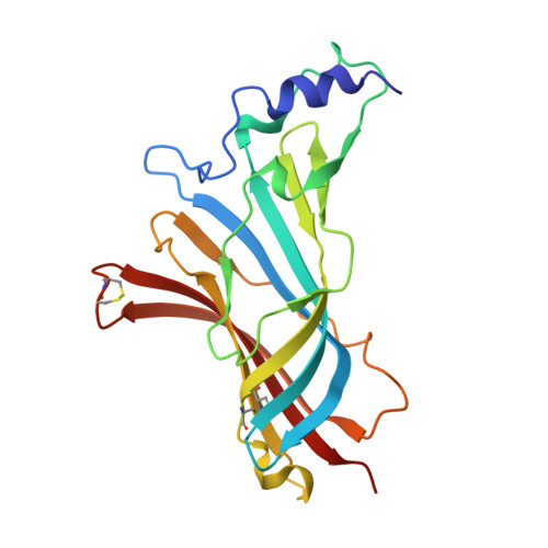
Ligand-binding domain of an alpha 7-nicotinic receptor chimera and its complex with agonist.

Li, S.X.Huang, S.Bren, N.Noridomi, K.Dellisanti, C.D.Sine, S.M.Chen, L.

(2011) Nat Neurosci 14: 1253-1259

  • DOI: https://doi.org/10.1038/nn.2908
  • Primary Citation of Related Structures:  
    3SQ6, 3SQ9

  • PubMed Abstract: 

    The α(7) acetylcholine receptor (AChR) mediates pre- and postsynaptic neurotransmission in the central nervous system and is a potential therapeutic target in neurodegenerative, neuropsychiatric and inflammatory disorders. We determined the crystal structure of the extracellular domain of a receptor chimera constructed from the human α(7) AChR and Lymnaea stagnalis acetylcholine binding protein (AChBP), which shares 64% sequence identity and 71% similarity with native α(7). We also determined the structure with bound epibatidine, a potent AChR agonist. Comparison of the structures revealed molecular rearrangements and interactions that mediate agonist recognition and early steps in signal transduction in α(7) AChRs. The structures further revealed a ring of negative charge within the central vestibule, poised to contribute to cation selectivity. Structure-guided mutational studies disclosed distinctive contributions to agonist recognition and signal transduction in α(7) AChRs. The structures provide a realistic template for structure-aided drug design and for defining structure-function relationships of α(7) AChRs.


  • Organizational Affiliation

    Molecular and Computational Biology, Department of Biological Sciences, University of Southern California, Los Angeles, California, USA.


Macromolecules
Find similar proteins by:  (by identity cutoff)  |  3D Structure
Entity ID: 1
MoleculeChains Sequence LengthOrganismDetailsImage
Neuronal acetylcholine receptor subunit alpha-7, Acetylcholine-binding protein
A, B, C, D, E
204Homo sapiensLymnaea stagnalis
This entity is chimeric
Mutation(s): 0 
Gene Names: CHRNA7
Entity Groups  
Sequence Clusters30% Identity50% Identity70% Identity90% Identity95% Identity100% Identity
Glycosylation
Glycosylation Sites: 2
Sequence Annotations
Expand
  • Reference Sequence
Oligosaccharides

Help

Entity ID: 2
MoleculeChains Length2D Diagram Glycosylation3D Interactions
2-acetamido-2-deoxy-beta-D-glucopyranose-(1-4)-2-acetamido-2-deoxy-beta-D-glucopyranose
K
2N-Glycosylation
Glycosylation Resources
GlyTouCan:  G42666HT
GlyCosmos:  G42666HT
GlyGen:  G42666HT
Small Molecules
Ligands 2 Unique
IDChains Name / Formula / InChI Key2D Diagram3D Interactions
NAG
Query on NAG

Download Ideal Coordinates CCD File 
BA [auth G]
DA [auth H]
EA [auth H]
GA [auth I]
M [auth A]
2-acetamido-2-deoxy-beta-D-glucopyranose
C8 H15 N O6
OVRNDRQMDRJTHS-FMDGEEDCSA-N
EPJ
Query on EPJ

Download Ideal Coordinates CCD File 
AA [auth G]
CA [auth H]
FA [auth I]
HA [auth J]
L [auth A]
EPIBATIDINE
C11 H13 Cl N2
NLPRAJRHRHZCQQ-IVZWLZJFSA-N
Experimental Data & Validation

Experimental Data

  • Method: X-RAY DIFFRACTION
  • Resolution: 2.80 Å
  • R-Value Free:  0.260 (Depositor), 0.244 (DCC) 
  • R-Value Work:  0.234 (Depositor), 0.220 (DCC) 
  • R-Value Observed: 0.234 (Depositor) 
Space Group: P 1 21 1
Unit Cell:
Length ( Å )Angle ( ˚ )
a = 81.237α = 90
b = 141.069β = 99.65
c = 130.207γ = 90
Software Package:
Software NamePurpose
DENZOdata reduction
SCALEPACKdata scaling
CNSrefinement
PDB_EXTRACTdata extraction
HKL-2000data scaling

Structure Validation

View Full Validation Report


Ligand Structure Quality Assessment 

Created with Raphaël 2.3.0Worse 01 BetterLigand structure goodness of fit to experimental dataBest fitted NAGClick on this verticalbar to view detailsBest fitted EPJClick on this verticalbar to view details

Entry History 

Deposition Data

Revision History  (Full details and data files)

  • Version 1.0: 2011-09-28
    Type: Initial release
  • Version 1.1: 2015-02-04
    Changes: Database references
  • Version 1.2: 2017-11-08
    Changes: Refinement description
  • Version 2.0: 2020-07-29
    Type: Remediation
    Reason: Carbohydrate remediation
    Changes: Atomic model, Data collection, Derived calculations, Structure summary
  • Version 2.1: 2024-10-09
    Changes: Data collection, Database references, Structure summary