3U9B | pdb_00003u9b

Structure of apo-CATI


Experimental Data Snapshot

  • Method: X-RAY DIFFRACTION
  • Resolution: 3.20 Å
  • R-Value Free: 
    0.301 (Depositor), 0.301 (DCC) 
  • R-Value Work: 
    0.235 (Depositor), 0.236 (DCC) 
  • R-Value Observed: 
    0.238 (Depositor) 

wwPDB Validation 3D Report Full Report

Validation slider image for 3U9B

This is version 1.3 of the entry. See complete history

Literature

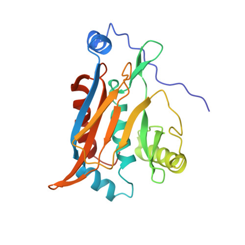
The structural basis for substrate versatility of chloramphenicol acetyltransferase CAT(I).

Biswas, T.Houghton, J.L.Garneau-Tsodikova, S.Tsodikov, O.V.

(2012) Protein Sci 21: 520-530

  • DOI: https://doi.org/10.1002/pro.2036
  • Primary Citation Related Structures: 
    3U9B, 3U9F

  • PubMed Abstract: 

    Novel antibiotics are needed to overcome the challenge of continually evolving bacterial resistance. This has led to a renewed interest in mechanistic studies of once popular antibiotics like chloramphenicol (CAM). Chloramphenicol acetyltransferases (CATs) are enzymes that covalently modify CAM, rendering it inactive against its target, the ribosome, and thereby causing resistance to CAM. Of the three major types of CAT (CAT(I-III)), the CAM-specific CAT(III) has been studied extensively. Much less is known about another clinically important type, CAT(I). In addition to inactivating CAM and unlike CAT(III), CAT(I) confers resistance to a structurally distinct antibiotic, fusidic acid. The origin of the broader substrate specificity of CAT(I) has not been fully elucidated. To understand the substrate binding features of CAT(I), its crystal structures in the unbound (apo) and CAM-bound forms were determined. The analysis of these and previously determined CAT(I)-FA and CAT(III)-CAM structures revealed interactions responsible for CAT(I) binding to its substrates and clarified the broader substrate preference of CAT(I) compared to that of CAT(III).


  • Organizational Affiliation
    • Department of Medicinal Chemistry, University of Michigan, Ann Arbor, Michigan 48109, USA.

Macromolecule Content 

  • Total Structure Weight: 231.23 kDa 
  • Atom Count: 15,981 
  • Modeled Residue Count: 1,930 
  • Deposited Residue Count: 1,971 
  • Unique protein chains: 1

Macromolecules

Find similar proteins by:|  3D Structure
Entity ID: 1
MoleculeChains  Sequence LengthOrganismDetailsImage
Chloramphenicol acetyltransferase
A, B, C, D, E
A, B, C, D, E, F, G, H, I
219Escherichia coliMutation(s): 0 
Gene Names: cat
EC: 2.3.1.28
UniProt
Find proteins for P62577 (Escherichia coli)
Explore P62577 
Go to UniProtKB:  P62577
Entity Groups
Sequence Clusters30% Identity50% Identity70% Identity90% Identity95% Identity100% Identity
UniProt GroupP62577
Sequence Annotations
Expand
Reference Sequence

Experimental Data & Validation

Experimental Data

  • Method: X-RAY DIFFRACTION
  • Resolution: 3.20 Å
  • R-Value Free:  0.301 (Depositor), 0.301 (DCC) 
  • R-Value Work:  0.235 (Depositor), 0.236 (DCC) 
  • R-Value Observed: 0.238 (Depositor) 
Space Group: P 1 21 1
Unit Cell:
Length ( Å )Angle ( ˚ )
a = 115.159α = 90
b = 102.743β = 119.94
c = 114.093γ = 90
Software Package:
Software NamePurpose
HKL-2000data collection
MOLREPphasing
REFMACrefinement
HKL-2000data reduction
HKL-2000data scaling

Structure Validation

View Full Validation Report


Entry History 

Deposition Data

Revision History  (Full details and data files)

  • Version 1.0: 2012-02-15
    Type: Initial release
  • Version 1.1: 2012-02-29
    Changes: Structure summary
  • Version 1.2: 2012-04-04
    Changes: Database references
  • Version 1.3: 2024-02-28
    Changes: Data collection, Database references