3UEL

Crystal structure of the catalytic domain of rat poly (ADP-ribose) glycohydrolase bound to ADP-HPD


Experimental Data Snapshot

  • Method: X-RAY DIFFRACTION
  • Resolution: 3.00 Å
  • R-Value Free: 
    0.274 (Depositor), 0.280 (DCC) 
  • R-Value Work: 
    0.245 (Depositor), 0.250 (DCC) 
  • R-Value Observed: 
    0.247 (Depositor) 

wwPDB Validation   3D Report Full Report


Ligand Structure Quality Assessment 

Created with Raphaël 2.3.0Worse 01 BetterLigand structure goodness of fit to experimental dataBest fitted A1RClick on this verticalbar to view details

This is version 1.2 of the entry. See complete history


Literature

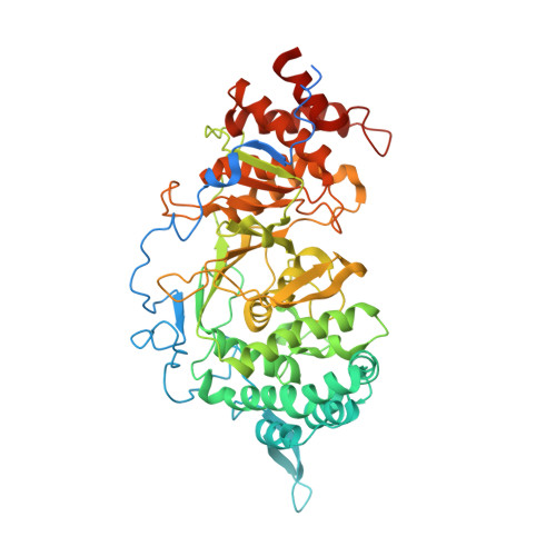
Structure of mammalian poly(ADP-ribose) glycohydrolase reveals a flexible tyrosine clasp as a substrate-binding element.

Kim, I.K.Kiefer, J.R.Ho, C.M.Stegeman, R.A.Classen, S.Tainer, J.A.Ellenberger, T.

(2012) Nat Struct Mol Biol 19: 653-656

  • DOI: https://doi.org/10.1038/nsmb.2305
  • Primary Citation of Related Structures:  
    3UEK, 3UEL

  • PubMed Abstract: 

    Reversible post-translational modification by poly(ADP-ribose) (PAR) regulates chromatin structure, DNA repair and cell fate in response to genotoxic stress. PAR glycohydrolase (PARG) removes PAR chains from poly ADP-ribosylated proteins to restore protein function and release oligo(ADP-ribose) chains to signal damage. Here we report crystal structures of mammalian PARG and its complex with a substrate mimic that reveal an open substrate-binding site and a unique 'tyrosine clasp' enabling endoglycosidic cleavage of branched PAR chains.


  • Organizational Affiliation

    Department of Biochemistry and Molecular Biophysics, Washington University School of Medicine, St. Louis, Missouri, USA.


Macromolecules
Find similar proteins by:  (by identity cutoff)  |  3D Structure
Entity ID: 1
MoleculeChains Sequence LengthOrganismDetailsImage
Poly(ADP-ribose) glycohydrolase
A, B, C
588Rattus norvegicusMutation(s): 0 
Gene Names: Parg
EC: 3.2.1.143
UniProt
Find proteins for Q9QYM2 (Rattus norvegicus)
Explore Q9QYM2 
Go to UniProtKB:  Q9QYM2
Entity Groups  
Sequence Clusters30% Identity50% Identity70% Identity90% Identity95% Identity100% Identity
UniProt GroupQ9QYM2
Sequence Annotations
Expand
  • Reference Sequence
Small Molecules
Binding Affinity Annotations 
IDSourceBinding Affinity
A1R BindingDB:  3UEL IC50: min: 100, max: 1400 (nM) from 5 assay(s)
Experimental Data & Validation

Experimental Data

  • Method: X-RAY DIFFRACTION
  • Resolution: 3.00 Å
  • R-Value Free:  0.274 (Depositor), 0.280 (DCC) 
  • R-Value Work:  0.245 (Depositor), 0.250 (DCC) 
  • R-Value Observed: 0.247 (Depositor) 
Space Group: C 2 2 21
Unit Cell:
Length ( Å )Angle ( ˚ )
a = 130.773α = 90
b = 195.989β = 90
c = 163.45γ = 90
Software Package:
Software NamePurpose
HKL-2000data collection
SOLVEphasing
REFMACrefinement
HKL-2000data reduction
SCALEPACKdata scaling

Structure Validation

View Full Validation Report


Ligand Structure Quality Assessment 

Created with Raphaël 2.3.0Worse 01 BetterLigand structure goodness of fit to experimental dataBest fitted A1RClick on this verticalbar to view details

Entry History 

Deposition Data

Revision History  (Full details and data files)

  • Version 1.0: 2012-05-23
    Type: Initial release
  • Version 1.1: 2012-07-18
    Changes: Database references
  • Version 1.2: 2024-02-28
    Changes: Data collection, Database references, Derived calculations