3UJL

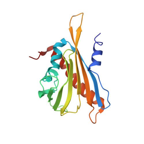
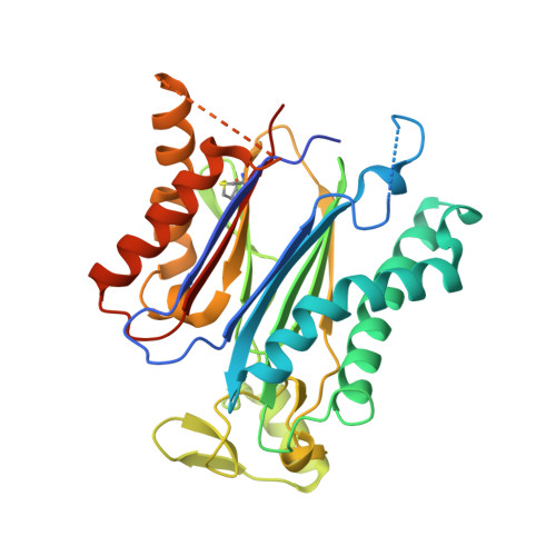
Crystal structure of abscisic acid bound PYL2 in complex with type 2C protein phosphatase ABI2


Experimental Data Snapshot

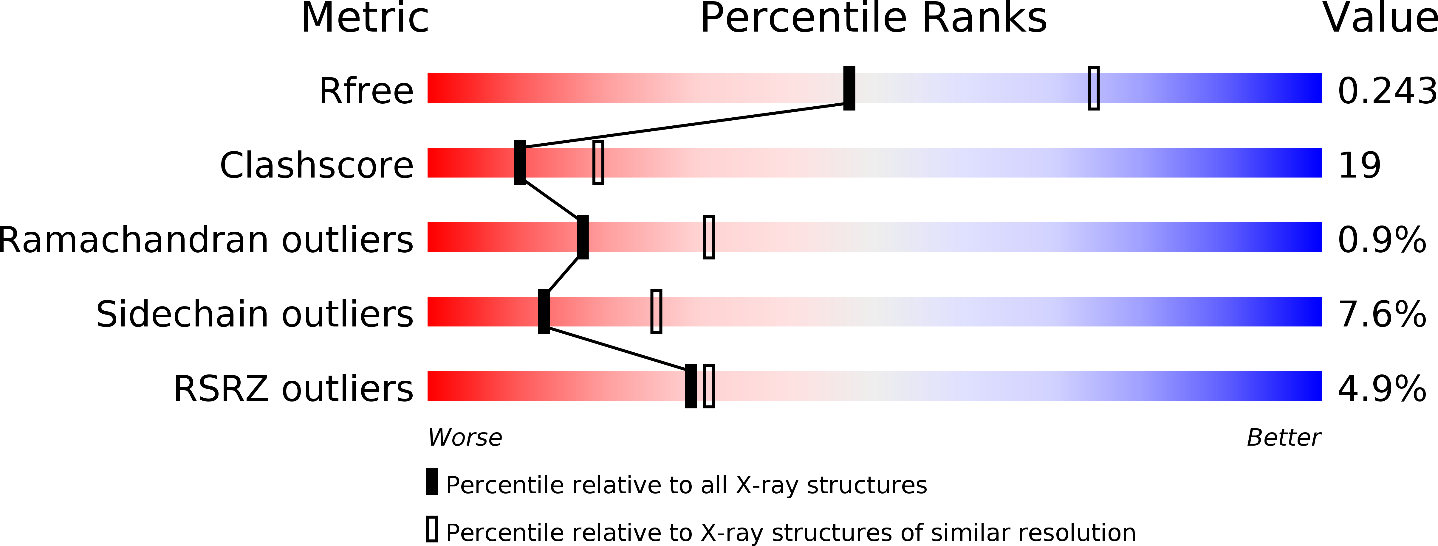
  • Method: X-RAY DIFFRACTION
  • Resolution: 2.50 Å
  • R-Value Free: 0.248 
  • R-Value Work: 0.223 
  • R-Value Observed: 0.224 

wwPDB Validation   3D Report Full Report


Ligand Structure Quality Assessment 


This is version 1.2 of the entry. See complete history


Literature

Molecular mimicry regulates ABA signaling by SnRK2 kinases and PP2C phosphatases.

Soon, F.F.Ng, L.M.Zhou, X.E.West, G.M.Kovach, A.Tan, M.H.Suino-Powell, K.M.He, Y.Xu, Y.Chalmers, M.J.Brunzelle, J.S.Zhang, H.Yang, H.Jiang, H.Li, J.Yong, E.L.Cutler, S.Zhu, J.K.Griffin, P.R.Melcher, K.Xu, H.E.

(2012) Science 335: 85-88

  • DOI: https://doi.org/10.1126/science.1215106
  • Primary Citation of Related Structures:  
    3UJG, 3UJK, 3UJL

  • PubMed Abstract: 

    Abscisic acid (ABA) is an essential hormone for plants to survive environmental stresses. At the center of the ABA signaling network is a subfamily of type 2C protein phosphatases (PP2Cs), which form exclusive interactions with ABA receptors and subfamily 2 Snfl-related kinase (SnRK2s). Here, we report a SnRK2-PP2C complex structure, which reveals marked similarity in PP2C recognition by SnRK2 and ABA receptors. In the complex, the kinase activation loop docks into the active site of PP2C, while the conserved ABA-sensing tryptophan of PP2C inserts into the kinase catalytic cleft, thus mimicking receptor-PP2C interactions. These structural results provide a simple mechanism that directly couples ABA binding to SnRK2 kinase activation and highlight a new paradigm of kinase-phosphatase regulation through mutual packing of their catalytic sites.


  • Organizational Affiliation

    Laboratory of Structural Sciences, Van Andel Research Institute, 333 Bostwick Avenue NE, Grand Rapids, MI 49503, USA.


Macromolecules
Find similar proteins by:  (by identity cutoff)  |  3D Structure
Entity ID: 1
MoleculeChains Sequence LengthOrganismDetailsImage
Abscisic acid receptor PYL2177Arabidopsis thalianaMutation(s): 0 
Gene Names: PYL2RCAR14At2g26040T19L18.15
UniProt
Find proteins for O80992 (Arabidopsis thaliana)
Explore O80992 
Go to UniProtKB:  O80992
Entity Groups  
Sequence Clusters30% Identity50% Identity70% Identity90% Identity95% Identity100% Identity
UniProt GroupO80992
Sequence Annotations
Expand
  • Reference Sequence
Find similar proteins by:  (by identity cutoff)  |  3D Structure
Entity ID: 2
MoleculeChains Sequence LengthOrganismDetailsImage
Protein phosphatase 2C 77324Arabidopsis thalianaMutation(s): 0 
Gene Names: ABI2At5g57050MHM17.19
EC: 3.1.3.16
UniProt
Find proteins for O04719 (Arabidopsis thaliana)
Explore O04719 
Go to UniProtKB:  O04719
Entity Groups  
Sequence Clusters30% Identity50% Identity70% Identity90% Identity95% Identity100% Identity
UniProt GroupO04719
Sequence Annotations
Expand
  • Reference Sequence
Experimental Data & Validation

Experimental Data

  • Method: X-RAY DIFFRACTION
  • Resolution: 2.50 Å
  • R-Value Free: 0.248 
  • R-Value Work: 0.223 
  • R-Value Observed: 0.224 
  • Space Group: P 21 21 21
Unit Cell:
Length ( Å )Angle ( ˚ )
a = 61.43α = 90
b = 98.587β = 90
c = 132.835γ = 90
Software Package:
Software NamePurpose
DENZOdata reduction
PHENIXrefinement
PDB_EXTRACTdata extraction
HKL-2000data collection
SCALEPACKdata scaling
PHASESphasing

Structure Validation

View Full Validation Report


Ligand Structure Quality Assessment 


Entry History 

Deposition Data

Revision History  (Full details and data files)

  • Version 1.0: 2012-02-15
    Type: Initial release
  • Version 1.1: 2012-03-28
    Changes: Source and taxonomy
  • Version 1.2: 2024-10-09
    Changes: Data collection, Database references, Derived calculations, Structure summary