3UOU | pdb_00003uou

Crystal structure of the Kunitz-type protease inhibitor ShPI-1 Lys13Leu mutant in complex with pancreatic elastase


Experimental Data Snapshot

  • Method: X-RAY DIFFRACTION
  • Resolution: 2.00 Å
  • R-Value Free: 
    0.211 (Depositor), 0.201 (DCC) 
  • R-Value Work: 
    0.166 (Depositor), 0.159 (DCC) 
  • R-Value Observed: 
    0.168 (Depositor) 

Starting Models: experimental
View more details

wwPDB Validation 3D Report Full Report

Validation slider image for 3UOU

This is version 1.4 of the entry. See complete history

Literature

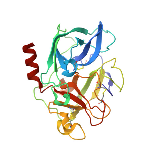
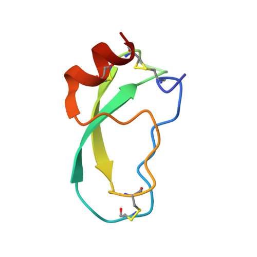
Three-dimensional Structure of a Kunitz-type Inhibitor in Complex with an Elastase-like Enzyme.

Garcia-Fernandez, R.Perbandt, M.Rehders, D.Ziegelmuller, P.Piganeau, N.Hahn, U.Betzel, C.Chavez, M.A.Redecke, L.

(2015) J Biological Chem 290: 14154-14165

  • DOI: https://doi.org/10.1074/jbc.M115.647586
  • Primary Citation Related Structures: 
    3UOU

  • PubMed Abstract: 

    Elastase-like enzymes are involved in important diseases such as acute pancreatitis, chronic inflammatory lung diseases, and cancer. Structural insights into their interaction with specific inhibitors will contribute to the development of novel anti-elastase compounds that resist rapid oxidation and proteolysis. Proteinaceous Kunitz-type inhibitors homologous to the bovine pancreatic trypsin inhibitor (BPTI) provide a suitable scaffold, but the structural aspects of their interaction with elastase-like enzymes have not been elucidated. Here, we increased the selectivity of ShPI-1, a versatile serine protease inhibitor from the sea anemone Stichodactyla helianthus with high biomedical and biotechnological potential, toward elastase-like enzymes by substitution of the P1 residue (Lys(13)) with leucine. The variant (rShPI-1/K13L) exhibits a novel anti-porcine pancreatic elastase (PPE) activity together with a significantly improved inhibition of human neuthrophil elastase and chymotrypsin. The crystal structure of the PPE·rShPI-1/K13L complex determined at 2.0 Å resolution provided the first details of the canonical interaction between a BPTI-Kunitz-type domain and elastase-like enzymes. In addition to the essential impact of the variant P1 residue for complex stability, the interface is improved by increased contributions of the primary and secondary binding loop as compared with similar trypsin and chymotrypsin complexes. A comparison of the interaction network with elastase complexes of canonical inhibitors from the chelonian in family supports a key role of the P3 site in ShPI-1 in directing its selectivity against pancreatic and neutrophil elastases. Our results provide the structural basis for site-specific mutagenesis to further improve the binding affinity and/or direct the selectivity of BPTI-Kunitz-type inhibitors toward elastase-like enzymes.


  • Organizational Affiliation
    • From the Centro de Estudio de Proteínas, Facultad de Biología, Universidad de la Habana, 20146 Habana, Cuba.

Macromolecule Content 

  • Total Structure Weight: 33.45 kDa 
  • Atom Count: 2,591 
  • Modeled Residue Count: 294 
  • Deposited Residue Count: 295 
  • Unique protein chains: 2

Macromolecules

Find similar proteins by:|  3D Structure
Entity ID: 1
MoleculeChains  Sequence LengthOrganismDetailsImage
Chymotrypsin-like elastase family member 1240Sus scrofaMutation(s): 0 
EC: 3.4.21.36
UniProt
Find proteins for P00772 (Sus scrofa)
Explore P00772 
Go to UniProtKB:  P00772
Entity Groups
Sequence Clusters30% Identity50% Identity70% Identity90% Identity95% Identity100% Identity
UniProt GroupP00772
Sequence Annotations
Expand
Reference Sequence
Find similar proteins by:|  3D Structure
Entity ID: 2
MoleculeChains  Sequence LengthOrganismDetailsImage
Kunitz-type proteinase inhibitor SHPI-155Stichodactyla helianthusMutation(s): 1 
UniProt
Find proteins for P31713 (Stichodactyla helianthus)
Explore P31713 
Go to UniProtKB:  P31713
Entity Groups
Sequence Clusters30% Identity50% Identity70% Identity90% Identity95% Identity100% Identity
UniProt GroupP31713
Sequence Annotations
Expand
Reference Sequence

Small Molecules

Ligands 2 Unique
IDChains Name / Formula / InChI Key2D Diagram3D Interactions
SO4

Query on SO4


Download:Ideal Coordinates CCD File
C [auth A]
D [auth A]
E [auth A]
F [auth A]
G [auth A]
C [auth A],
D [auth A],
E [auth A],
F [auth A],
G [auth A],
H [auth A],
O [auth B],
P [auth B]
SULFATE ION
O4 S
QAOWNCQODCNURD-UHFFFAOYSA-L
GOL

Query on GOL


Download:Ideal Coordinates CCD File
I [auth A]
J [auth A]
K [auth A]
L [auth A]
M [auth A]
I [auth A],
J [auth A],
K [auth A],
L [auth A],
M [auth A],
N [auth A],
Q [auth B]
GLYCEROL
C3 H8 O3
PEDCQBHIVMGVHV-UHFFFAOYSA-N

Experimental Data & Validation

Experimental Data

  • Method: X-RAY DIFFRACTION
  • Resolution: 2.00 Å
  • R-Value Free:  0.211 (Depositor), 0.201 (DCC) 
  • R-Value Work:  0.166 (Depositor), 0.159 (DCC) 
  • R-Value Observed: 0.168 (Depositor) 
Space Group: C 1 2 1
Unit Cell:
Length ( Å )Angle ( ˚ )
a = 132.75α = 90
b = 47.18β = 100.07
c = 42.68γ = 90
Software Package:
Software NamePurpose
DNAdata collection
PHASERphasing
PHENIXrefinement
MOSFLMdata reduction
SCALAdata scaling

Structure Validation

View Full Validation Report


Entry History 

Deposition Data

Revision History  (Full details and data files)

  • Version 1.0: 2012-11-21
    Type: Initial release
  • Version 1.1: 2015-04-29
    Changes: Database references
  • Version 1.2: 2015-06-17
    Changes: Database references
  • Version 1.3: 2023-09-13
    Changes: Data collection, Database references, Derived calculations, Refinement description
  • Version 1.4: 2024-11-20
    Changes: Structure summary