3VXV | pdb_00003vxv

Crystal structure of methyl CpG Binding Domain of MBD4 in complex with the 5mCG/TG sequence


Experimental Data Snapshot

  • Method: X-RAY DIFFRACTION
  • Resolution: 2.00 Å
  • R-Value Free: 
    0.224 (Depositor), 0.214 (DCC) 
  • R-Value Work: 
    0.188 (Depositor), 0.177 (DCC) 
  • R-Value Observed: 
    0.189 (Depositor) 

wwPDB Validation 3D Report Full Report

Validation slider image for 3VXV

This is version 1.2 of the entry. See complete history

Literature

Structural basis of the versatile DNA recognition ability of the methyl-CpG binding domain of methyl-CpG binding domain protein 4

Otani, J.Arita, K.Kato, T.Kinoshita, M.Kimura, H.Suetake, I.Tajima, S.Ariyoshi, M.Shirakawa, M.

(2013) J Biological Chem 288: 6351-6362

  • DOI: https://doi.org/10.1074/jbc.M112.431098
  • Primary Citation Related Structures: 
    3VXV, 3VXX, 3VYB, 3VYQ

  • PubMed Abstract: 

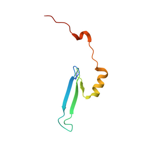
    The methyl-CpG binding domain (MBD) protein MBD4 participates in DNA repair as a glycosylase that excises mismatched thymine bases in CpG sites and also functions in transcriptional repression. Unlike other MBD proteins, MBD4 recognizes not only methylated CpG dinucleotides ((5m)CG/(5m)CG) but also T/G mismatched sites generated by spontaneous deamination of 5-methylcytosine ((5m)CG/TG). The glycosylase activity of MBD4 is also implicated in active DNA demethylation initiated by the deaminase-catalyzed conversion of 5-methylcytosine to thymine. Here, we report the crystal structures of the MBD of MBD4 (MBDMBD4) complexed with (5m)CG/(5m)CG and (5m)CG/TG. The crystal structures show that the DNA interface of MBD4 has flexible structural features and harbors an extensive water network that supports its dual base specificities. Combined with the results of biochemical analyses, the crystal structure of MBD4 bound to 5-hydroxymethylcytosine further demonstrates that MBDMBD4 is able to recognize a wide range of 5-methylcytosine modifications through the unique water network. The versatile base recognition ability of MBDMBD4 implies multifunctional roles for MBD4 in the regulation of dynamic DNA methylation patterns coupled with deamination and/or oxidation of 5-methylcytosine.


  • Organizational Affiliation
    • Graduate School of Engineering, Kyoto University, Kyoto 615-8510, Japan.

Macromolecule Content 

  • Total Structure Weight: 16.97 kDa 
  • Atom Count: 1,285 
  • Modeled Residue Count: 93 
  • Deposited Residue Count: 97 
  • Unique protein chains: 1
  • Unique nucleic acid chains: 2

Macromolecules


Find similar proteins by:|  3D Structure
Entity ID: 1
MoleculeChains  Sequence LengthOrganismDetailsImage
Methyl-CpG-binding domain protein 469Mus musculusMutation(s): 0 
Gene Names: Mbd4
EC: 3.2.2
UniProt
Find proteins for Q9Z2D7 (Mus musculus)
Explore Q9Z2D7 
Go to UniProtKB:  Q9Z2D7
Entity Groups
Sequence Clusters30% Identity50% Identity70% Identity90% Identity95% Identity100% Identity
UniProt GroupQ9Z2D7
Sequence Annotations
Expand
Reference Sequence
Find similar nucleic acids by:  Sequence
Entity ID: 2
MoleculeChains LengthOrganismImage
DNA (5'-D(*GP*TP*CP*AP*CP*TP*AP*CP*(5CM)P*GP*GP*AP*CP*A)-3')14N/A
Sequence Annotations
Expand
Reference Sequence
Find similar nucleic acids by:  Sequence
Entity ID: 3
MoleculeChains LengthOrganismImage
DNA (5'-D(*GP*TP*CP*TP*GP*GP*TP*AP*GP*TP*GP*AP*CP*T)-3')14N/A
Sequence Annotations
Expand
Reference Sequence

Small Molecules

Ligands 2 Unique
IDChains Name / Formula / InChI Key2D Diagram3D Interactions
EDO

Query on EDO


Download:Ideal Coordinates CCD File
F [auth A]
G [auth A]
H [auth A]
I [auth A]
J [auth A]
F [auth A],
G [auth A],
H [auth A],
I [auth A],
J [auth A],
L [auth C]
1,2-ETHANEDIOL
C2 H6 O2
LYCAIKOWRPUZTN-UHFFFAOYSA-N
ACT

Query on ACT


Download:Ideal Coordinates CCD File
D [auth A],
E [auth A],
K [auth B]
ACETATE ION
C2 H3 O2
QTBSBXVTEAMEQO-UHFFFAOYSA-M

Experimental Data & Validation

Experimental Data

  • Method: X-RAY DIFFRACTION
  • Resolution: 2.00 Å
  • R-Value Free:  0.224 (Depositor), 0.214 (DCC) 
  • R-Value Work:  0.188 (Depositor), 0.177 (DCC) 
  • R-Value Observed: 0.189 (Depositor) 
Space Group: C 2 2 21
Unit Cell:
Length ( Å )Angle ( ˚ )
a = 89.074α = 90
b = 94.989β = 90
c = 54.738γ = 90
Software Package:
Software NamePurpose
HKL-2000data collection
SOLVEphasing
PHENIXrefinement
HKL-2000data reduction
HKL-2000data scaling

Structure Validation

View Full Validation Report


Entry History 

Deposition Data

Revision History  (Full details and data files)

  • Version 1.0: 2013-01-16
    Type: Initial release
  • Version 1.1: 2013-08-14
    Changes: Database references
  • Version 1.2: 2024-11-06
    Changes: Data collection, Database references, Derived calculations, Structure summary