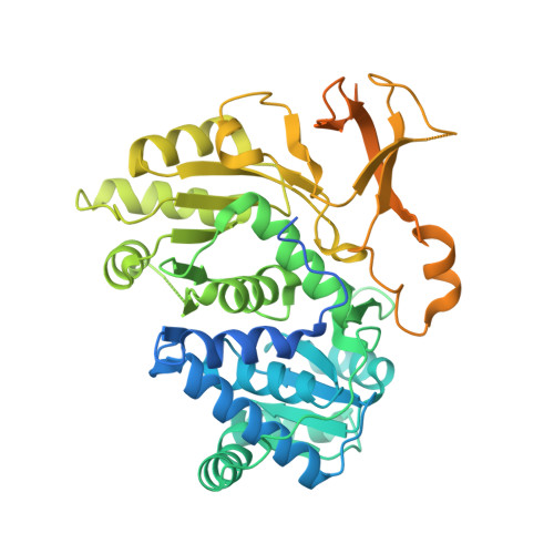
The crystal structure of the adenylation enzyme VinN reveals a unique beta-amino acid recognition mechanism
Miyanaga, A., Cieslak, J., Shinohara, Y., Kudo, F., Eguchi, T.(2014) J Biological Chem 289: 31448-31457
- PubMed: 25246523
- DOI: https://doi.org/10.1074/jbc.M114.602326
- Primary Citation of Related Structures:
3WV4, 3WV5, 3WVN - PubMed Abstract:
Adenylation enzymes play important roles in the biosynthesis and degradation of primary and secondary metabolites. Mechanistic insights into the recognition of α-amino acid substrates have been obtained for α-amino acid adenylation enzymes. The Asp residue is invariant and is essential for the stabilization of the α-amino group of the substrate. In contrast, the β-amino acid recognition mechanism of adenylation enzymes is still unclear despite the importance of β-amino acid activation for the biosynthesis of various natural products. Herein, we report the crystal structure of the stand-alone adenylation enzyme VinN, which specifically activates (2S,3S)-3-methylaspartate (3-MeAsp) in vicenistatin biosynthesis. VinN has an overall structure similar to that of other adenylation enzymes. The structure of the complex with 3-MeAsp revealed that a conserved Asp(230) residue is used in the recognition of the β-amino group of 3-MeAsp similar to α-amino acid adenylation enzymes. A mutational analysis and structural comparison with α-amino acid adenylation enzymes showed that the substrate-binding pocket of VinN has a unique architecture to accommodate 3-MeAsp as a β-amino acid substrate. Thus, the VinN structure allows the first visualization of the interaction of an adenylation enzyme with a β-amino acid and provides new mechanistic insights into the selective recognition of β-amino acids in this family of enzymes.
- From the Departments of Chemistry and.
Organizational Affiliation:


