3ZO9

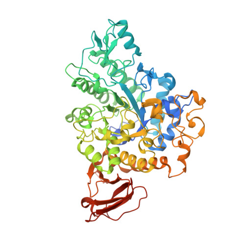
The structure of Trehalose Synthase (TreS) of Mycobacterium smegmatis


Experimental Data Snapshot

  • Method: X-RAY DIFFRACTION
  • Resolution: 1.84 Å
  • R-Value Free: 0.195 
  • R-Value Work: 0.163 
  • R-Value Observed: 0.164 

Starting Model: experimental
View more details

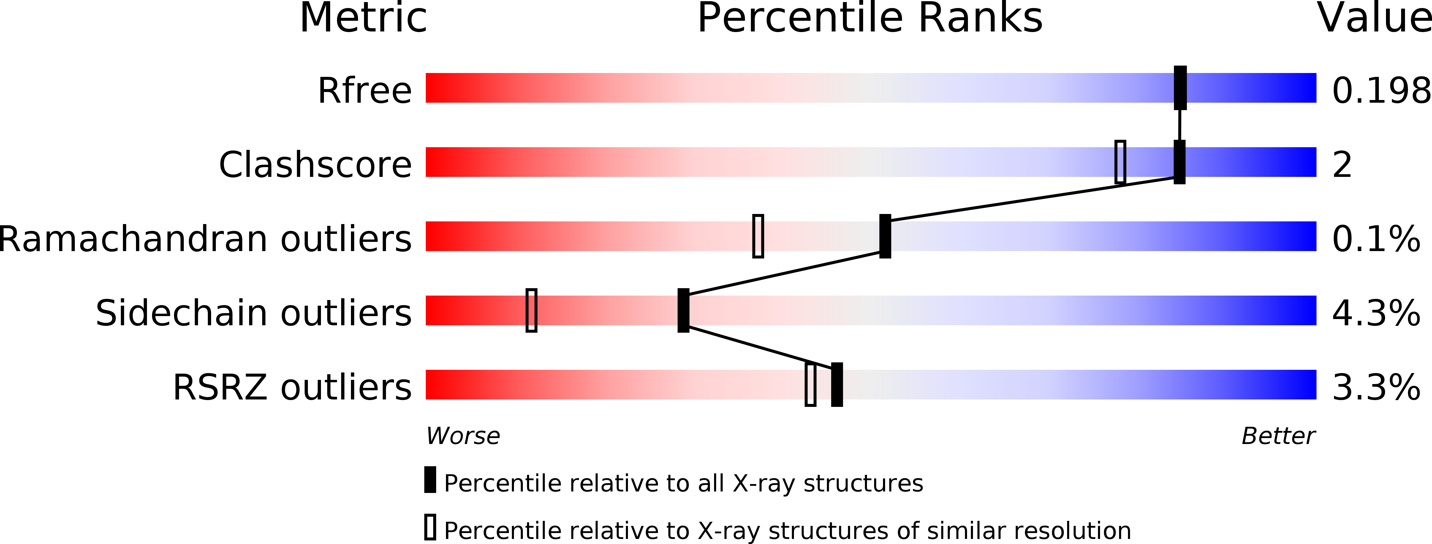
wwPDB Validation   3D Report Full Report


This is version 1.2 of the entry. See complete history


Literature

The Structure of the Mycobacterium Smegmatis Trehalose Synthase Reveals an Unusual Active Site Configuration and Acarbose-Binding Mode.

Caner, S.Nguyen, N.Aguda, A.Zhang, R.Pan, Y.T.Withers, S.G.Brayer, G.D.

(2013) Glycobiology 23: 1075

  • DOI: https://doi.org/10.1093/glycob/cwt044
  • Primary Citation of Related Structures:  
    3ZO9, 3ZOA

  • PubMed Abstract: 

    Trehalose synthase (TreS) catalyzes the reversible conversion of maltose into trehalose in mycobacteria as one of three biosynthetic pathways to this nonreducing disaccharide. Given the importance of trehalose to survival of mycobacteria, there has been considerable interest in understanding the enzymes involved in its production; indeed the structures of the key enzymes in the other two pathways have already been determined. Herein, we present the first structure of TreS from Mycobacterium smegmatis, thereby providing insights into the catalytic machinery involved in this intriguing intramolecular reaction. This structure, which is of interest both mechanistically and as a potential pharmaceutical target, reveals a narrow and enclosed active site pocket within which intramolecular substrate rearrangements can occur. We also present the structure of a complex of TreS with acarbose, revealing a hitherto unsuspected oligosaccharide-binding site within the C-terminal domain. This may well provide an anchor point for the association of TreS with glycogen, thereby enhancing its role in glycogen biosynthesis and degradation.


  • Organizational Affiliation

    Department of Biochemistry and Molecular Biology, University of British Columbia, British Columbia, Canada.


Macromolecules
Find similar proteins by:  (by identity cutoff)  |  3D Structure
Entity ID: 1
MoleculeChains Sequence LengthOrganismDetailsImage
TREHALOSE SYNTHASE/AMYLASE TRES
A, B
593Mycolicibacterium smegmatisMutation(s): 0 
EC: 3.2.1.1 (PDB Primary Data), 5.4.99.16 (PDB Primary Data)
UniProt
Find proteins for A0R6E0 (Mycolicibacterium smegmatis (strain ATCC 700084 / mc(2)155))
Explore A0R6E0 
Go to UniProtKB:  A0R6E0
Entity Groups  
Sequence Clusters30% Identity50% Identity70% Identity90% Identity95% Identity100% Identity
UniProt GroupA0R6E0
Sequence Annotations
Expand
  • Reference Sequence
Small Molecules
Ligands 3 Unique
IDChains Name / Formula / InChI Key2D Diagram3D Interactions
CA
Query on CA

Download Ideal Coordinates CCD File 
E [auth A],
K [auth B]
CALCIUM ION
Ca
BHPQYMZQTOCNFJ-UHFFFAOYSA-N
CL
Query on CL

Download Ideal Coordinates CCD File 
C [auth A]
D [auth A]
F [auth A]
G [auth A]
I [auth B]
C [auth A],
D [auth A],
F [auth A],
G [auth A],
I [auth B],
J [auth B],
L [auth B]
CHLORIDE ION
Cl
VEXZGXHMUGYJMC-UHFFFAOYSA-M
MG
Query on MG

Download Ideal Coordinates CCD File 
H [auth A],
M [auth B]
MAGNESIUM ION
Mg
JLVVSXFLKOJNIY-UHFFFAOYSA-N
Experimental Data & Validation

Experimental Data

  • Method: X-RAY DIFFRACTION
  • Resolution: 1.84 Å
  • R-Value Free: 0.195 
  • R-Value Work: 0.163 
  • R-Value Observed: 0.164 
  • Space Group: I 41
Unit Cell:
Length ( Å )Angle ( ˚ )
a = 127.55α = 90
b = 127.55β = 90
c = 217.76γ = 90
Software Package:
Software NamePurpose
PHENIXrefinement
XDSdata reduction
XDSdata scaling
PHASERphasing

Structure Validation

View Full Validation Report


Entry History 

Deposition Data

Revision History  (Full details and data files)

  • Version 1.0: 2013-07-17
    Type: Initial release
  • Version 1.1: 2013-08-07
    Changes: Database references
  • Version 1.2: 2023-12-20
    Changes: Data collection, Database references, Derived calculations, Other, Refinement description