4A6D | pdb_00004a6d

Crystal structure of human N-acetylserotonin methyltransferase (ASMT) in complex with SAM


Experimental Data Snapshot

  • Method: X-RAY DIFFRACTION
  • Resolution: 2.40 Å
  • R-Value Free: 
    0.204 (Depositor), 0.210 (DCC) 
  • R-Value Work: 
    0.176 (Depositor), 0.187 (DCC) 
  • R-Value Observed: 
    0.177 (Depositor) 

wwPDB Validation 3D Report Full Report

Validation slider image for 4A6D

Ligand Structure Quality Assessment 


This is version 1.2 of the entry. See complete history

Literature

Crystal Structure and Functional Mapping of Human Asmt, the Last Enzyme of the Melatonin Synthesis Pathway.

Botros, H.G.Legrand, P.Pagan, C.Bondet, V.Weber, P.Ben-Abdallah, M.Lemiere, N.Huguet, G.Bellalou, J.Maronde, E.Beguin, P.Haouz, A.Shepard, W.Bourgeron, T.

(2013) J Pineal Res 54: 46

  • DOI: https://doi.org/10.1111/j.1600-079X.2012.01020.x
  • Primary Citation Related Structures: 
    4A6D, 4A6E

  • PubMed Abstract: 

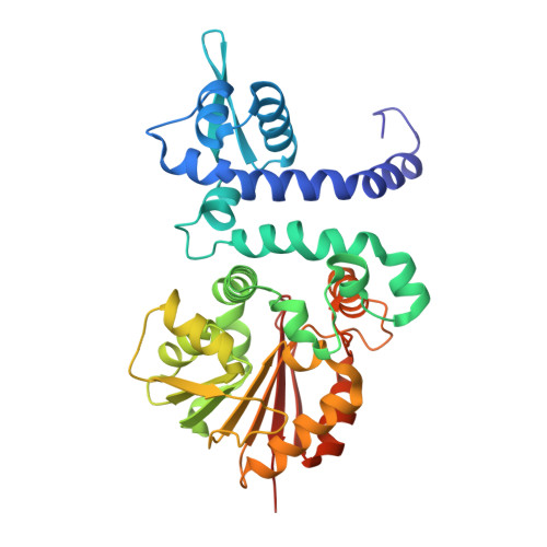
    Melatonin is a synchronizer of many physiological processes. Abnormal melatonin signaling is associated with human disorders related to sleep, metabolism, and neurodevelopment. Here, we present the X-ray crystal structure of human N-acetyl serotonin methyltransferase (ASMT), the last enzyme of the melatonin biosynthesis pathway. The polypeptide chain of ASMT consists of a C-terminal domain, which is typical of other SAM-dependent O-methyltransferases, and an N-terminal domain, which intertwines several helices with another monomer to form the physiologically active dimer. Using radioenzymology, we analyzed 20 nonsynonymous variants identified through the 1000 genomes project and in patients with neuropsychiatric disorders. We found that the majority of these mutations reduced or abolished ASMT activity including one relatively frequent polymorphism in the Han Chinese population (N17K, rs17149149). Overall, we estimate that the allelic frequency of ASMT deleterious mutations ranges from 0.66% in Europe to 2.97% in Asia. Mapping of the variants on to the 3-dimensional structure clarifies why some are harmful and provides a structural basis for understanding melatonin deficiency in humans.


  • Organizational Affiliation
    • Institut Pasteur, Human Genetics and Cognitive Functions Unit, Paris, France CNRS URA 2182 'Genes, synapses and cognition', Institut Pasteur, Paris, France University Paris Diderot, Sorbonne Paris Cité, Human Genetics and Cognitive Functions, Paris, France Synchrotron SOLEIL, L'Orme des Merisiers, Saint Aubin BP48, Gif-sur-Yvette, France Institut Pasteur, Plate forme 5, 25 rue Dr. Roux, Paris, France Institut Pasteur, Plate forme 6, CNRS-UMR3528, 25 rue Dr. Roux, Paris, France Institute for Anatomy III, Goethe University, Frankfurt, Germany.

Macromolecule Content 

  • Total Structure Weight: 40.48 kDa 
  • Atom Count: 2,834 
  • Modeled Residue Count: 345 
  • Deposited Residue Count: 353 
  • Unique protein chains: 1

Macromolecules

Find similar proteins by:|  3D Structure
Entity ID: 1
MoleculeChains  Sequence LengthOrganismDetailsImage
HYDROXYINDOLE O-METHYLTRANSFERASE353Homo sapiensMutation(s): 0 
EC: 2.1.1.4
UniProt & NIH Common Fund Data Resources
Find proteins for P46597 (Homo sapiens)
Explore P46597 
Go to UniProtKB:  P46597
PHAROS:  P46597
GTEx:  ENSG00000196433 
Entity Groups
Sequence Clusters30% Identity50% Identity70% Identity90% Identity95% Identity100% Identity
UniProt GroupP46597
Sequence Annotations
Expand
Reference Sequence

Small Molecules

Ligands 4 Unique
IDChains Name / Formula / InChI Key2D Diagram3D Interactions
SAM

Query on SAM


Download:Ideal Coordinates CCD File
D [auth A]S-ADENOSYLMETHIONINE
C15 H22 N6 O5 S
MEFKEPWMEQBLKI-FCKMPRQPSA-N
SO4

Query on SO4


Download:Ideal Coordinates CCD File
E [auth A],
F [auth A],
G [auth A],
H [auth A]
SULFATE ION
O4 S
QAOWNCQODCNURD-UHFFFAOYSA-L
GOL

Query on GOL


Download:Ideal Coordinates CCD File
I [auth A]GLYCEROL
C3 H8 O3
PEDCQBHIVMGVHV-UHFFFAOYSA-N
ZN

Query on ZN


Download:Ideal Coordinates CCD File
B [auth A],
C [auth A]
ZINC ION
Zn
PTFCDOFLOPIGGS-UHFFFAOYSA-N

Experimental Data & Validation

Experimental Data

  • Method: X-RAY DIFFRACTION
  • Resolution: 2.40 Å
  • R-Value Free:  0.204 (Depositor), 0.210 (DCC) 
  • R-Value Work:  0.176 (Depositor), 0.187 (DCC) 
  • R-Value Observed: 0.177 (Depositor) 
Space Group: H 3 2
Unit Cell:
Length ( Å )Angle ( ˚ )
a = 170.4α = 90
b = 170.4β = 90
c = 123.32γ = 120
Software Package:
Software NamePurpose
BUSTERrefinement
XDSdata reduction
XSCALEdata scaling

Structure Validation

View Full Validation Report


Ligand Structure Quality Assessment 


Entry History 

Deposition Data

Revision History  (Full details and data files)

  • Version 1.0: 2012-11-07
    Type: Initial release
  • Version 1.1: 2013-08-28
    Changes: Database references
  • Version 1.2: 2024-05-08
    Changes: Data collection, Database references, Derived calculations, Other