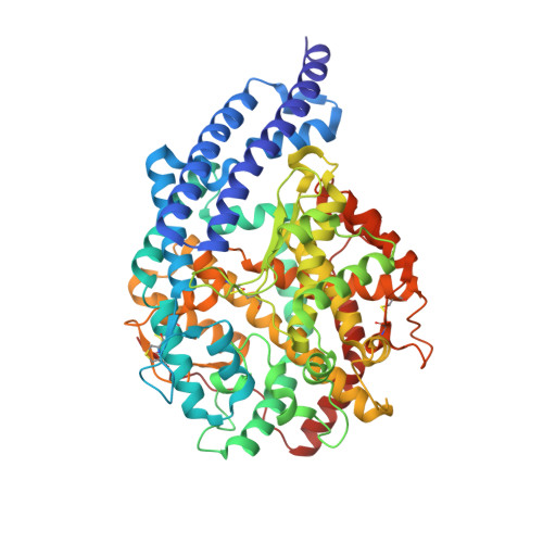
4AA2

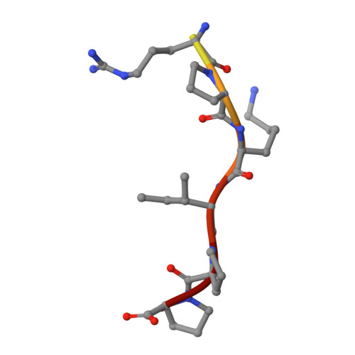
Crystal structure of ANCE in complex with bradykinin potentiating peptide b


Experimental Data Snapshot

  • Method: X-RAY DIFFRACTION
  • Resolution: 1.99 Å
  • R-Value Free: 0.226 
  • R-Value Work: 0.203 
  • R-Value Observed: 0.204 

Starting Model: experimental
View more details

wwPDB Validation   3D Report Full Report


Ligand Structure Quality Assessment 


This is version 2.2 of the entry. See complete history


Literature

Structural Basis of Peptide Recognition by the Angiotensin-I Converting Enzyme Homologue Ance from Drosophila Melanogaster

Akif, M.Masuyer, G.Bingham, R.J.Sturrock, E.D.Isaac, R.E.Acharya, K.R.

(2012) FEBS J 279: 4525

  • DOI: https://doi.org/10.1111/febs.12038
  • Primary Citation of Related Structures:  
    4AA1, 4AA2, 4ASQ, 4ASR

  • PubMed Abstract: 

    Human somatic angiotensin-1 converting enzyme (ACE) is a zinc-dependent exopeptidase, that catalyses the conversion of the decapeptide angiotensin I to the octapeptide angiotensin II, by removing a C-terminal dipeptide. It is the principal component of the renin-angiotensin-aldosterone system that regulates blood pressure. Hence it is an important therapeutic target for the treatment of hypertension and cardiovascular disorders. Here, we report the structures of an ACE homologue from Drosophila melanogaster (AnCE; a proven structural model for the more complex human ACE) co-crystallized with mammalian peptide substrates (bradykinin, Thr(6) -bradykinin, angiotensin I and a snake venom peptide inhibitor, bradykinin-potentiating peptide-b). The structures determined at 2-Å resolution illustrate that both angiotensin II (the cleaved product of angiotensin I by AnCE) and bradykinin-potentiating peptide-b bind in an analogous fashion at the active site of AnCE, but also exhibit significant differences. In addition, the binding of Arg-Pro-Pro, the cleavage product of bradykinin and Thr(6) - bradykinin, provides additional detail of the general peptide binding in AnCE. Thus the new structures of AnCE complexes presented here improves our understanding of the binding of peptides and the mechanism by which peptides inhibit this family of enzymes. The atomic coordinates and structure factors for AnCE-Ang II (code 4AA1), AnCE-BPPb (code 4AA2), AnCE-BK (code 4ASQ) and AnCE-Thr6-BK (code 4ASR) complexes have been deposited in the Protein Data Bank, Research Collaboratory for Structural Bioinformatics, Rutgers University, New Brunswick, NJ (http://www.rcsb.org/) • AnCE cleaves Ang I by enzymatic study (View interaction) • Bradykinin and AnCE bind by x-ray crystallography (View interaction) • BPP and AnCE bind by x-ray crystallography (View interaction) • AnCE cleaves Bradykinin by enzymatic study (View interaction) • Ang II and AnCE bind by x-ray crystallography (View interaction).


  • Organizational Affiliation

    Department of Biology and Biochemistry, University of Bath, UK.


Macromolecules
Find similar proteins by:  (by identity cutoff)  |  3D Structure
Entity ID: 1
MoleculeChains Sequence LengthOrganismDetailsImage
ANGIOTENSIN-CONVERTING ENZYME598Drosophila melanogasterMutation(s): 0 
EC: 3.4.15.1
UniProt
Find proteins for Q10714 (Drosophila melanogaster)
Explore Q10714 
Go to UniProtKB:  Q10714
Entity Groups  
Sequence Clusters30% Identity50% Identity70% Identity90% Identity95% Identity100% Identity
UniProt GroupQ10714
Glycosylation
Glycosylation Sites: 1Go to GlyGen: Q10714-1
Sequence Annotations
Expand
  • Reference Sequence

Find similar proteins by:  Sequence   |   3D Structure  

Entity ID: 2
MoleculeChains Sequence LengthOrganismDetailsImage
BRADYKININ-POTENTIATING PEPTIDE BB [auth P]11Gloydius blomhoffiiMutation(s): 0 
UniProt
Find proteins for P01021 (Gloydius blomhoffii)
Explore P01021 
Go to UniProtKB:  P01021
Entity Groups  
Sequence Clusters30% Identity50% Identity70% Identity90% Identity95% Identity100% Identity
UniProt GroupP01021
Sequence Annotations
Expand
  • Reference Sequence
Oligosaccharides

Help

Entity ID: 3
MoleculeChains Length2D Diagram Glycosylation3D Interactions
beta-D-mannopyranose-(1-6)-alpha-D-mannopyranose-(1-3)-[alpha-D-mannopyranose-(1-6)]beta-D-mannopyranose-(1-4)-2-acetamido-2-deoxy-beta-D-glucopyranose-(1-4)-2-acetamido-2-deoxy-beta-D-glucopyranoseC [auth B]6N-Glycosylation
Glycosylation Resources
GlyTouCan:  G64803HZ
GlyCosmos:  G64803HZ
GlyGen:  G64803HZ
Experimental Data & Validation

Experimental Data

  • Method: X-RAY DIFFRACTION
  • Resolution: 1.99 Å
  • R-Value Free: 0.226 
  • R-Value Work: 0.203 
  • R-Value Observed: 0.204 
  • Space Group: H 3
Unit Cell:
Length ( Å )Angle ( ˚ )
a = 173.214α = 90
b = 173.214β = 90
c = 102.898γ = 120
Software Package:
Software NamePurpose
REFMACrefinement
HKL-2000data reduction
SCALEPACKdata scaling
PHASERphasing

Structure Validation

View Full Validation Report


Ligand Structure Quality Assessment 


Entry History 

Deposition Data

Revision History  (Full details and data files)

  • Version 1.0: 2012-10-31
    Type: Initial release
  • Version 1.1: 2012-12-05
    Changes: Database references
  • Version 1.2: 2012-12-19
    Changes: Database references
  • Version 2.0: 2020-07-29
    Type: Remediation
    Reason: Carbohydrate remediation
    Changes: Advisory, Atomic model, Data collection, Derived calculations, Other, Structure summary
  • Version 2.1: 2023-12-20
    Changes: Data collection, Database references, Refinement description, Structure summary
  • Version 2.2: 2024-11-20
    Changes: Structure summary